
Patient guide · 01—06
Patient information
Understand your neurovascular care.
Clear explanations of diagnostic angiography, stroke treatment and prevention, treatment of aneurysms and vascular malformations, and care in a dedicated stroke unit.
Safety first
Risks shared by catheter-based procedures
Every procedure is planned individually. The likelihood and seriousness of complications depend on the condition being treated, urgency, anatomy, age, other illnesses and medicines.
- 01
Bruising, pain, bleeding, haematoma, blockage or injury at the wrist or groin access site; rarely, repair or transfusion may be required.
- 02
Damage, spasm, dissection or perforation of an artery; clot formation or material travelling downstream may cause a transient neurological deficit or stroke.
- 03
Allergic reaction to iodinated contrast and temporary worsening of kidney function, particularly when kidney disease, dehydration or diabetes is present.
- 04
Radiation exposure is kept as low as reasonably achievable; pregnancy or possible pregnancy must be discussed before non-emergency procedures.
- 05
Sedation or general anaesthesia may cause breathing, blood-pressure, heart-rhythm or medication-related complications.
- 06
Infection is uncommon. Any procedure involving the brain circulation can very rarely cause severe disability or death.
Your team reduces risk through imaging review, blood tests, medicine checks, sterile technique, continuous monitoring and a treatment plan tailored to you. Never stop prescribed antiplatelet or anticoagulant medication unless your treating team tells you exactly when and how.
Cerebral angiography
A precise map of the arteries and veins of the brain, head and neck.
01Diagnostic procedure
Cerebral angiography (DSA)
Open detailed guideClose detailed guide
Diagnostic procedure
Cerebral angiography (DSA)
Open detailed guideClose detailed guide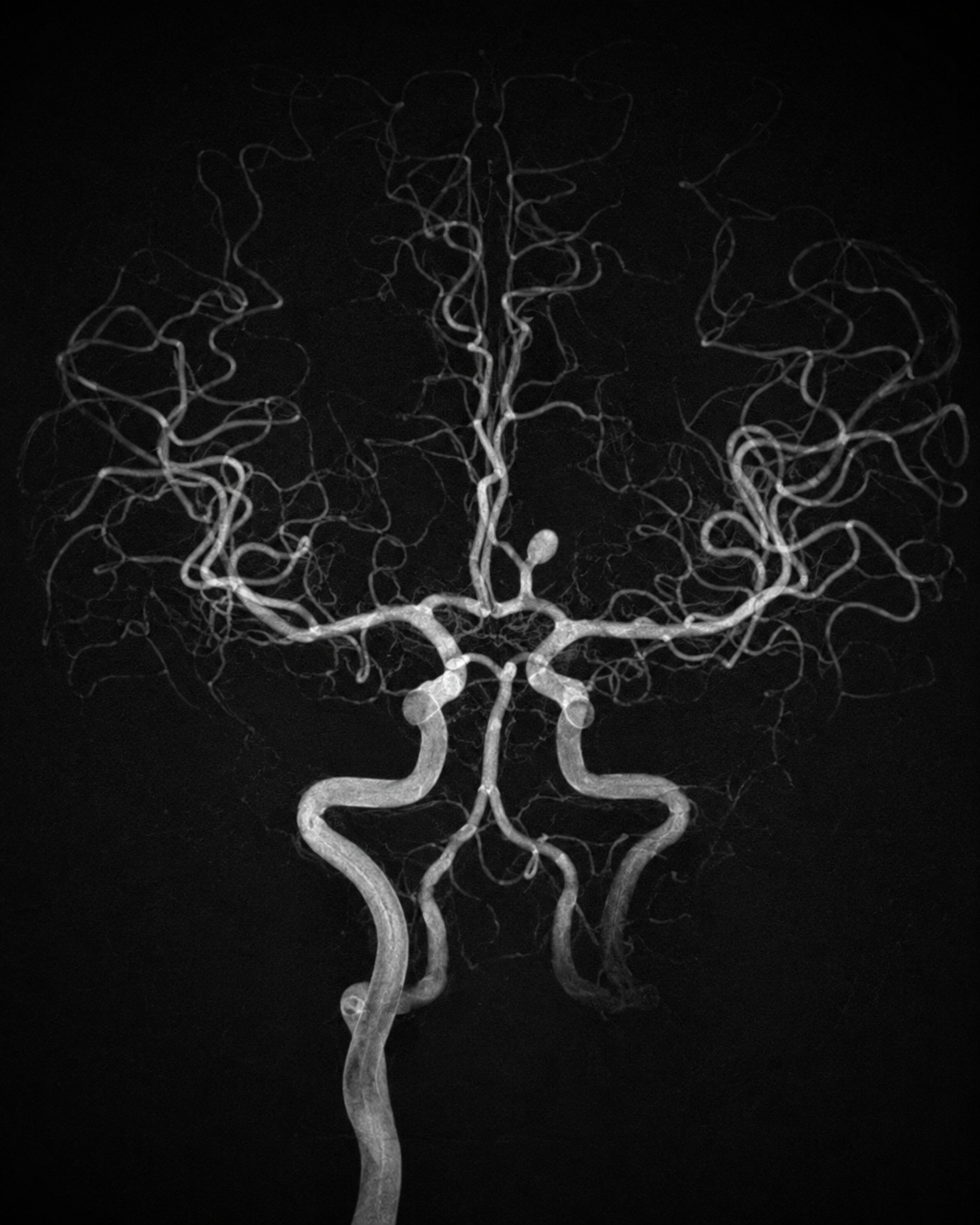
What it is and why it may be advised
Digital subtraction angiography uses a thin catheter, X-rays and iodinated contrast to show blood vessels in greater detail than CTA or MRA. It may be needed to define an aneurysm, AVM, fistula, stenosis, dissection or another vascular problem and to plan treatment.
How the strategy is chosen
The neurointerventional specialist first reviews CT, MRI, CTA or MRA. DSA is recommended only when the extra detail is expected to change diagnosis, risk assessment or treatment. The safest access route, vessels to study, need for 3D imaging and level of sedation are chosen for the individual patient.
What happens step by step
- 1
Beforehand: the team reviews allergies, kidney function, blood count, clotting, pregnancy possibility and all medicines. Fasting instructions depend on the planned sedation.
- 2
Preparation: monitoring is attached and the wrist or groin is cleaned and numbed. Most adults remain awake with local anaesthetic; light sedation may be offered.
- 3
Catheter navigation: a small sheath enters the artery. Under live X-ray guidance, a thin catheter is moved to selected neck vessels; it does not enter the brain itself.
- 4
Imaging: contrast is injected while rapid images are taken. Brief warmth, a metallic taste or flashing lights may occur. Additional angled or 3D runs may be needed.
- 5
Completion: the catheter and sheath are removed, pressure or a closure device seals the artery, and neurological observations and access-site checks follow.
Options and alternatives
- CTA or MRA may answer some questions without an arterial catheter, but may not provide the same spatial, flow or time-resolved detail.
- A diagnostic study may occasionally proceed directly to treatment, but only if this possibility was discussed and appropriate consent and preparation are in place.
- For non-urgent conditions, postponement or imaging surveillance may be reasonable when the expected information will not alter care.
Procedure-specific risks and complications
- Temporary symptoms such as headache, weakness, speech or visual disturbance; permanent neurological injury or stroke is uncommon but possible.
- Groin or wrist bleeding, arterial blockage, pseudoaneurysm or damage requiring additional treatment.
- Contrast allergy, nausea or kidney injury; the team may use hydration or adapt contrast use when risk is increased.
- Rare catheter-related vessel injury, dissection, perforation or intracranial bleeding.
After the procedure
- Observation usually lasts several hours. Keep the access limb still for the period advised and drink fluids if permitted.
- Mild bruising is common. Follow instructions about driving, lifting, bathing and restarting medicines.
- Seek urgent help for new neurological symptoms, severe headache, increasing swelling or bleeding, a cold/pale limb, chest pain or breathing difficulty.
Acute ischaemic stroke procedures
Emergency treatment aims to restore blood flow before more brain tissue is permanently injured.
01Emergency clot removal
Mechanical thrombectomy
Open detailed guideClose detailed guide
Emergency clot removal
Mechanical thrombectomy
Open detailed guideClose detailed guide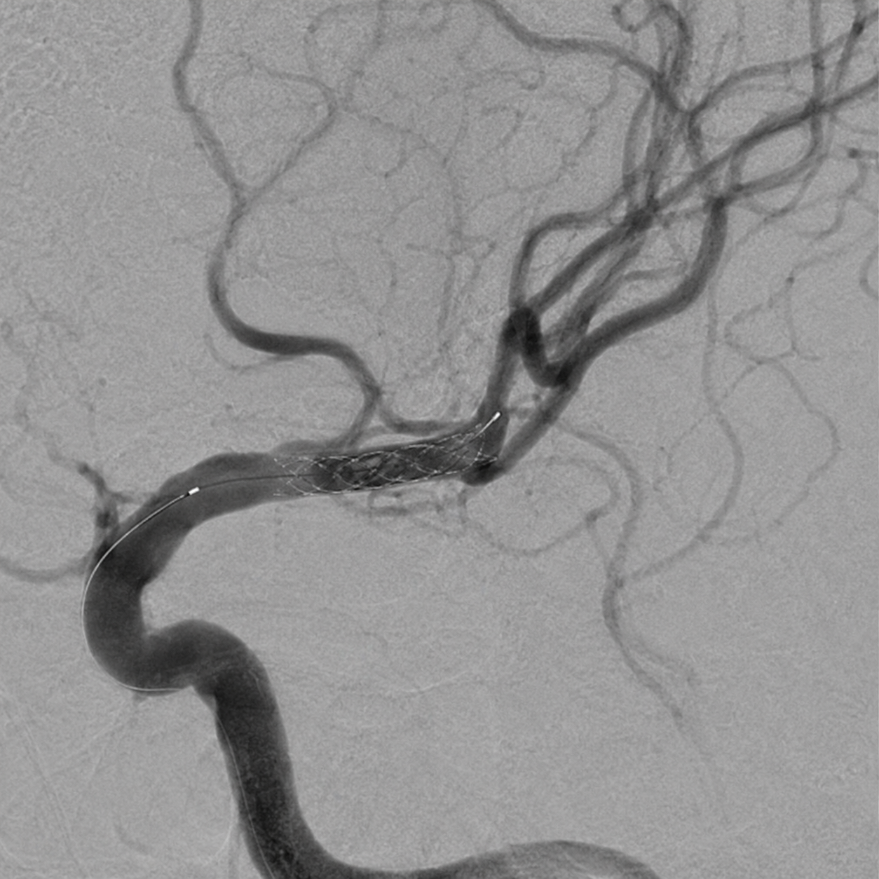
What it is and why it may be advised
Mechanical thrombectomy is an emergency catheter procedure that removes a clot from a large brain artery. It can substantially improve the chance of independence in appropriately selected patients, but it cannot reverse brain tissue that is already permanently damaged.
How the strategy is chosen
The stroke team combines symptom time, neurological examination, CT or MRI, vessel imaging and sometimes perfusion imaging. Treatment may be offered beyond the traditional early window in selected patients. If intravenous thrombolysis is suitable, it is usually given promptly and does not delay thrombectomy.
What happens step by step
- 1
Stroke-code assessment: immediate brain imaging excludes haemorrhage and identifies the blocked artery and tissue that may still be saved.
- 2
Preparation: the team checks medical history, medicines and blood tests while anaesthesia and the angiography team prepare in parallel.
- 3
Access and navigation: through the groin or wrist, a catheter is guided to the artery supplying the brain. Sedation or general anaesthesia is selected according to safety and clinical status.
- 4
Clot removal: aspiration, a stent retriever, or both are used to capture and remove the clot. More than one attempt or technique may be necessary.
- 5
Reperfusion check: angiography confirms the degree of restored flow and looks for an underlying narrowing. The access site is closed and the patient transfers to a stroke unit or intensive care area.
Options and alternatives
- Aspiration thrombectomy, stent-retriever thrombectomy, or a combined technique; device choice depends on the vessel and clot.
- Intravenous thrombolysis when eligible, alongside thrombectomy rather than as a reason to delay it.
- Best medical and supportive care when imaging shows no treatable large-vessel occlusion or when the expected procedural risk outweighs benefit.
Procedure-specific risks and complications
- Bleeding into or around the brain, including haemorrhagic transformation of the stroke.
- Failure to reopen the artery, incomplete reperfusion, re-occlusion, clot migration or a new stroke in another territory.
- Vessel spasm, dissection or perforation; emergency additional treatment or surgery may be required.
- Brain swelling, seizures, aspiration, infection and complications related to severe stroke may occur despite technically successful treatment.
After the procedure
- Frequent neurological, blood-pressure and access-site checks are performed; repeat CT or MRI is often arranged.
- Swallowing is checked before food, drink or tablets. Rehabilitation starts early when clinically safe.
- Recovery varies widely. The team explains the final reperfusion result, brain injury seen on follow-up imaging and the prevention plan.
02Bailout treatment
Rescue intracranial stenting
Open detailed guideClose detailed guide
Bailout treatment
Rescue intracranial stenting
Open detailed guideClose detailed guide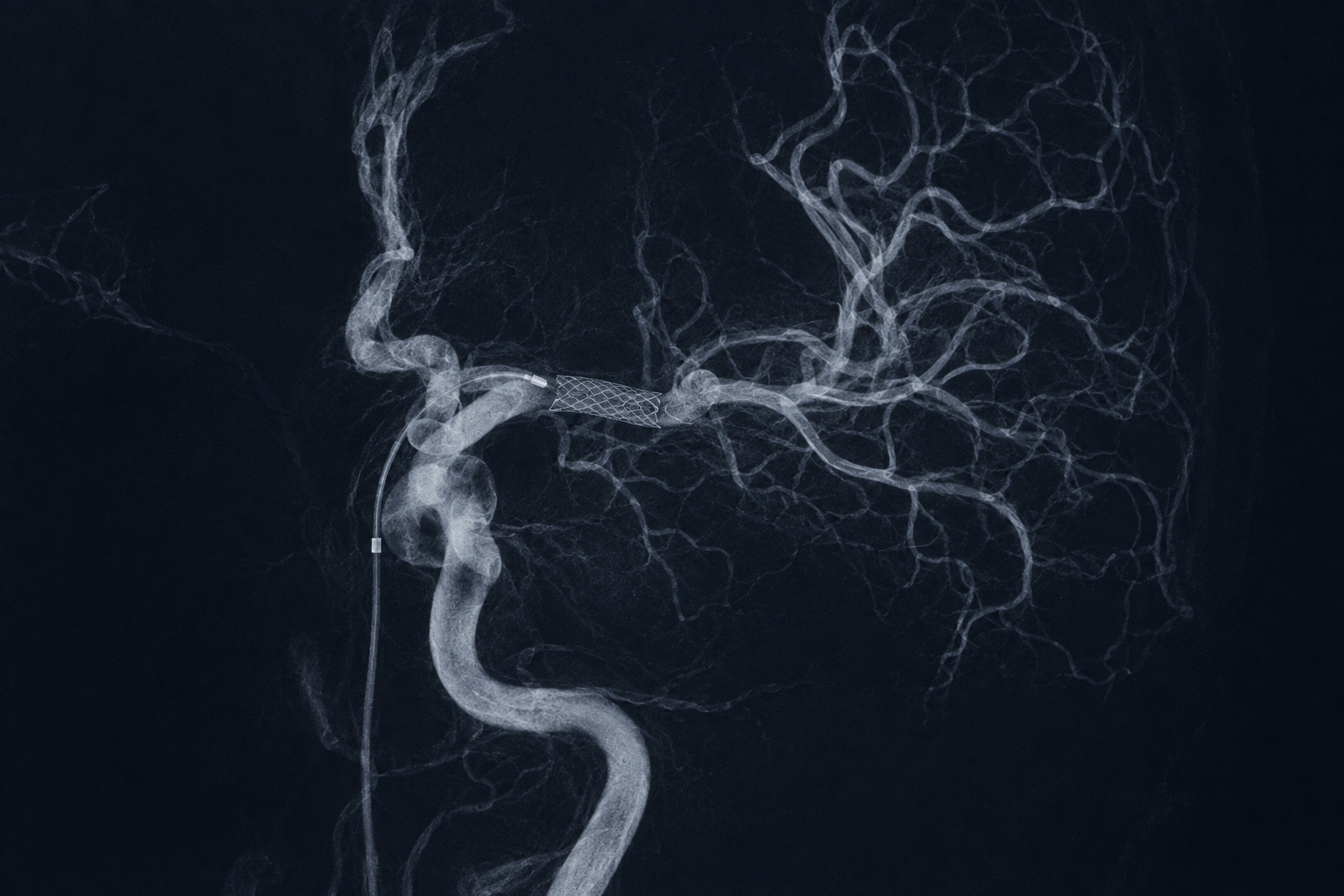
What it is and why it may be advised
Rescue stenting may be considered during thrombectomy when an artery repeatedly closes, a severe fixed narrowing prevents durable flow, or standard clot-removal techniques do not achieve adequate reperfusion. It is not required in most thrombectomies.
How the strategy is chosen
The decision is made in real time after angiography. The specialist weighs the amount of brain that may still be saved against bleeding risk, the need for urgent antiplatelet medication, the location and cause of narrowing, and alternatives such as further aspiration, angioplasty or stopping the procedure.
What happens step by step
- 1
Angiography after clot removal identifies persistent severe narrowing or repeated re-occlusion.
- 2
The team confirms that more blood flow is likely to benefit viable brain and considers the recent use of thrombolytic or anticoagulant medicines.
- 3
A microcatheter and wire cross the diseased segment. Gentle balloon angioplasty may be performed first in selected cases.
- 4
A self-expanding stent is deployed to support the artery. Antiplatelet medication is administered using a protocol adapted to bleeding risk.
- 5
Repeated images confirm flow and exclude perforation or immediate thrombosis, followed by close stroke-unit or intensive-care monitoring.
Options and alternatives
- Further thrombectomy or aspiration attempts when a residual clot is the main problem.
- Balloon angioplasty without a stent, short-acting antiplatelet medication, or observation if flow is acceptable.
- Stopping when further intervention is judged more dangerous than the likely benefit.
Procedure-specific risks and complications
- Intracranial haemorrhage, particularly because antiplatelet treatment may be necessary soon after a stroke or thrombolysis.
- Acute clotting inside the stent, later re-narrowing or re-occlusion, with recurrent stroke.
- Perforator-artery occlusion, vessel dissection or perforation and injury to nearby brain tissue.
- Need for prolonged dual-antiplatelet therapy, which increases bleeding risk and must not be interrupted without specialist advice.
After the procedure
- More intensive neurological and blood-pressure monitoring and early follow-up imaging are required.
- Antiplatelet medication is prescribed according to the device and bleeding risk; exact timing and duration are individual.
- Follow-up vascular imaging checks stent patency. New stroke symptoms or medication-related bleeding require urgent assessment.
Stroke prevention procedures
Procedures are only one part of prevention; risk-factor treatment remains essential before and after intervention.
01Cervical artery treatment
Carotid stenting & systemic atherosclerosis
Open detailed guideClose detailed guide
Cervical artery treatment
Carotid stenting & systemic atherosclerosis
Open detailed guideClose detailed guide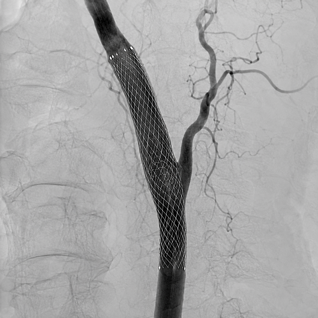
What it is and why it may be advised
Carotid artery stenting opens a significant narrowing in the neck artery that supplies the brain. It may reduce future stroke risk in selected symptomatic or high-risk patients, but it does not treat atherosclerosis throughout the body.
How the strategy is chosen
The choice between optimal medical therapy, carotid endarterectomy and carotid stenting is made after confirming the degree of stenosis, recent symptoms, plaque and arch anatomy, age, other illnesses and procedural risk. A multidisciplinary discussion is particularly important because surgery may be safer for some patients and stenting for others.
What happens step by step
- 1
Planning: ultrasound and CTA or MRA define the narrowing; cardiac risk, kidney function and medicines are reviewed. Prescribed antiplatelet treatment begins before elective stenting.
- 2
Protection: through the groin or wrist, a catheter reaches the carotid artery. A filter or flow-reversal system may be used to reduce debris reaching the brain.
- 3
Opening the artery: a balloon may gently widen the narrowing, then a self-expanding stent is positioned across the plaque.
- 4
Completion: further ballooning is used only if needed. Angiography checks brain flow and excludes immediate complications.
- 5
Monitoring: heart rate, blood pressure, neurological status and the access site are observed closely, often overnight.
Options and alternatives
- Best medical treatment: antiplatelet therapy when prescribed, intensive cholesterol reduction, blood-pressure and diabetes control, smoking cessation, exercise, diet and weight management.
- Carotid endarterectomy: surgical removal of plaque, often the preferred revascularisation option for suitable patients with recent symptoms.
- Carotid stenting: considered when anatomy, medical risk, prior surgery/radiation or other factors favour an endovascular approach.
Procedure-specific risks and complications
- Stroke or transient ischaemic attack from plaque or clot travelling to the brain during or after the procedure.
- Slow heart rate or low blood pressure from pressure on the carotid sinus; medication or temporary support may be needed.
- Bleeding, arterial injury, contrast/kidney complications and, rarely, excessive brain blood flow causing headache, seizures or haemorrhage.
- Clotting within the stent or later restenosis; surveillance and reliable antiplatelet use are important.
After the procedure
- Continue antiplatelet and cholesterol-lowering treatment exactly as prescribed and attend ultrasound or vascular follow-up.
- The wider disease still requires treatment: stop smoking, control blood pressure, cholesterol and diabetes, remain active and adopt a heart-healthy diet.
- Seek emergency help for any new facial droop, weakness, speech/vision difficulty, severe headache, chest pain or access-site bleeding.
02Selected ICAD treatment
Intracranial stenting & ICAD
Open detailed guideClose detailed guide
Selected ICAD treatment
Intracranial stenting & ICAD
Open detailed guideClose detailed guide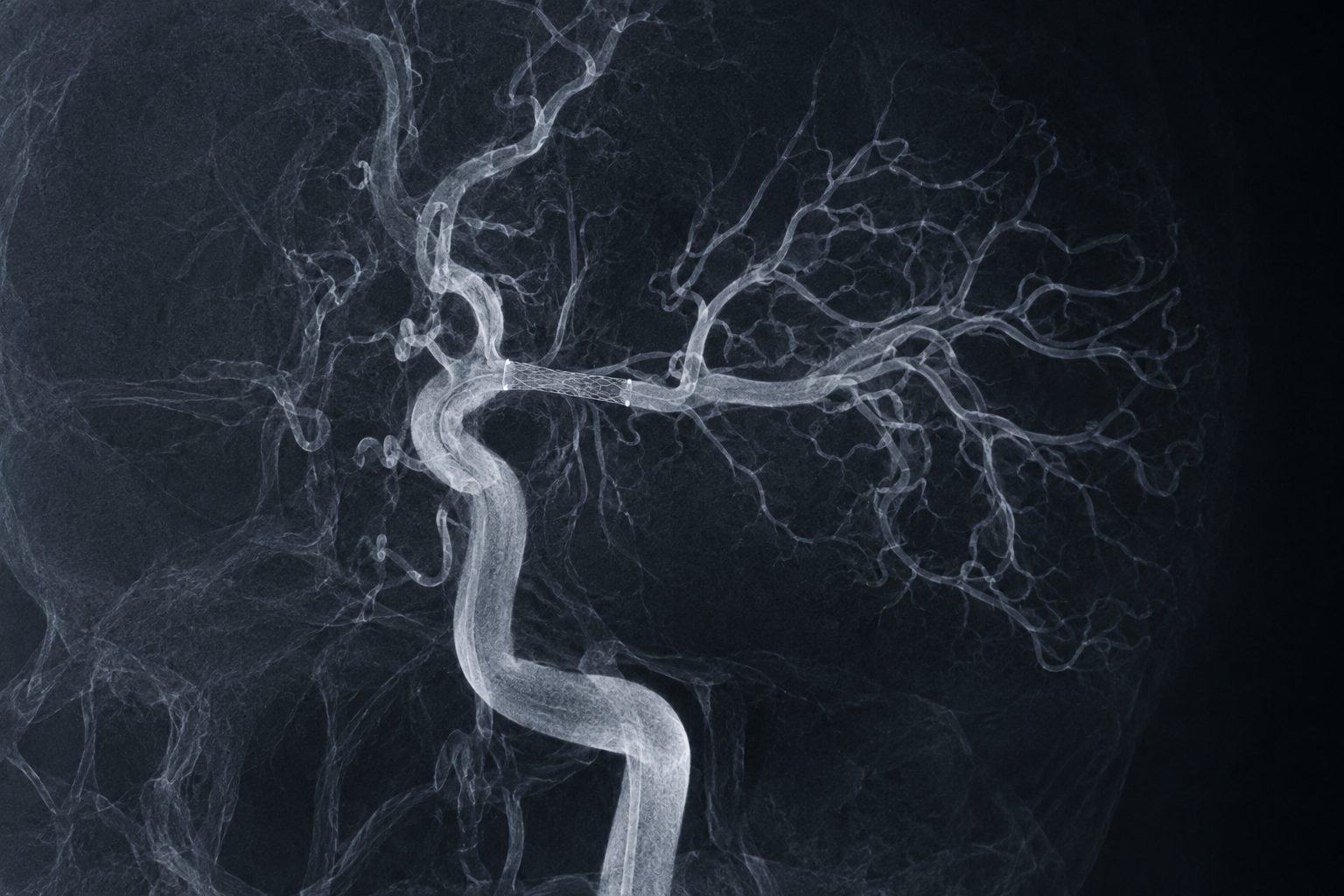
What it is and why it may be advised
Intracranial atherosclerotic disease (ICAD) is plaque-related narrowing of an artery inside the skull. For most patients, the first treatment is intensive medical therapy rather than a stent because early stenting can carry substantial stroke and bleeding risk.
How the strategy is chosen
The team confirms that symptoms arise from the narrowed artery, checks severity and blood-flow reserve, and reviews adherence to medical therapy. Intracranial angioplasty or stenting is reserved for carefully selected patients with recurrent ischaemia despite optimised treatment, usually after multidisciplinary review and when the acute phase has passed.
What happens step by step
- 1
Assessment: CTA, MRA, MRI/perfusion and sometimes DSA define the stenosis, collateral circulation and previous infarcts.
- 2
Medical optimisation: antiplatelet therapy, intensive lipid and blood-pressure management, diabetes care, smoking cessation and activity are addressed first.
- 3
If intervention is selected, dual-antiplatelet response, anatomy and device sizing are checked and the procedure is performed under close anaesthetic monitoring.
- 4
A microcatheter crosses the stenosis; cautious balloon angioplasty may be followed by a self-expanding or balloon-mounted stent in selected anatomy.
- 5
Final angiography and close post-procedure monitoring look for perforator occlusion, bleeding, clotting or excessive pressure changes.
Options and alternatives
- Aggressive medical management is standard first-line care for most symptomatic ICAD.
- Balloon angioplasty alone or stenting may be considered only in selected refractory cases at experienced centres.
- Bypass surgery is not routine stroke-prevention treatment for typical ICAD; clinical trials may be available for uncertain situations.
Procedure-specific risks and complications
- Stroke from perforator blockage, clot, plaque displacement, dissection or acute stent thrombosis.
- Intracranial bleeding from vessel injury or excessive reperfusion; neurological deterioration may be severe.
- Restenosis or late stent occlusion, requiring imaging surveillance and sometimes further treatment.
- Bleeding from necessary antiplatelet medication and the shared catheter, contrast and anaesthesia risks.
After the procedure
- Neurological and blood-pressure monitoring is intensive, with early brain imaging when indicated.
- Dual-antiplatelet therapy must be taken exactly as directed; missed doses can cause dangerous stent thrombosis.
- Long-term success depends on medical therapy and risk-factor control even when the artery has been opened.
Haemorrhagic stroke & vascular lesions
Treatment aims to stop or prevent bleeding while protecting normal brain vessels and tissue.
01Aneurysm treatment
Cerebral aneurysm embolisation
Open detailed guideClose detailed guide
Aneurysm treatment
Cerebral aneurysm embolisation
Open detailed guideClose detailed guide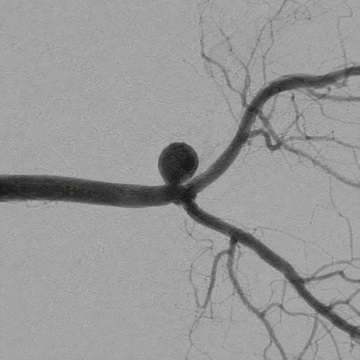
What it is and why it may be advised
An aneurysm is a weakened, bulging area of an artery. Endovascular treatment closes the aneurysm from inside the vessel or diverts blood away from it to reduce rupture or rebleeding risk.
How the strategy is chosen
Treatment is not automatic for every unruptured aneurysm. A multidisciplinary team weighs rupture history and estimated future risk against treatment risk, considering size, shape, growth, location, age, health and patient preference. A ruptured aneurysm is an emergency and is generally secured as early as feasible.
What happens step by step
- 1
Planning angiography defines the aneurysm neck, branches and access route. Medicines and anaesthesia are tailored to rupture status and the likely device.
- 2
A catheter is guided from the wrist or groin into the artery carrying the aneurysm under general anaesthesia in most treatments.
- 3
A microcatheter reaches the aneurysm or parent artery. Coils, an intrasaccular device, a supporting stent or a flow diverter may be deployed according to anatomy.
- 4
Angiography checks aneurysm exclusion, branch patency and any clot or vessel injury. Additional medication or devices may be required.
- 5
The patient transfers to a stroke unit or intensive care setting; ruptured aneurysms require broader subarachnoid-haemorrhage care.
Options and alternatives
- Imaging surveillance with risk-factor control for selected unruptured aneurysms when treatment risk is not lower than expected rupture risk.
- Endovascular options include simple or balloon-assisted coiling, stent-assisted coiling, intrasaccular flow disruption and flow diversion.
- Microsurgical clipping remains an important alternative and may be preferred for specific anatomy, patient factors or associated brain haemorrhage.
Procedure-specific risks and complications
- Clot formation, branch occlusion or embolism causing transient or permanent stroke.
- Aneurysm or artery perforation causing subarachnoid or intracerebral haemorrhage; emergency surgery may be required.
- Incomplete occlusion, recurrence or coil compaction requiring imaging follow-up and sometimes retreatment.
- Stents and flow diverters require antiplatelet medication, creating both clotting risk if interrupted and bleeding risk while taken.
After the procedure
- Uncomplicated elective treatment often requires short inpatient observation; recovery after rupture is longer and determined mainly by the original haemorrhage.
- Follow-up MRA, CTA or DSA checks durable occlusion. Keep all imaging and medication appointments.
- A sudden severe headache, new neurological deficit, seizure, collapse or unexpected bleeding requires emergency assessment.
02Abnormal vascular connections
AVM & dural AVF therapy
Open detailed guideClose detailed guide
Abnormal vascular connections
AVM & dural AVF therapy
Open detailed guideClose detailed guide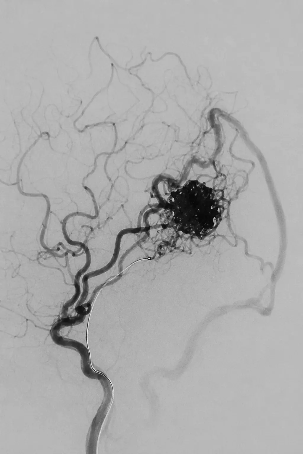
What it is and why it may be advised
A brain arteriovenous malformation (AVM) is a network of abnormal vessels connecting arteries and veins. A dural arteriovenous fistula (dAVF) is an abnormal direct connection within the covering of the brain. Their behaviour and bleeding risk vary greatly, especially according to venous drainage.
How the strategy is chosen
DSA maps feeding arteries, the AVM nidus or fistula point, draining veins and high-risk features. A multidisciplinary team considers symptoms, previous haemorrhage, location, size, venous drainage and treatment risk. The goal may be complete cure, staged risk reduction or treatment of a dangerous weak point.
What happens step by step
- 1
Detailed MRI/CT and DSA define the anatomy and whether urgent treatment is needed.
- 2
Under general anaesthesia, a microcatheter is guided through an artery—and for some dAVFs through a vein—to the abnormal connection.
- 3
Liquid embolic material, coils or both are delivered in a controlled way while protecting normal arteries and veins.
- 4
Treatment may be staged. Completion angiography checks residual shunting and normal brain circulation.
- 5
Follow-up imaging is essential because a residual or recurrent connection may need further embolisation, surgery or radiosurgery.
Options and alternatives
- Observation with imaging may be appropriate for selected lower-risk lesions, especially when treatment risk exceeds natural-history risk.
- Endovascular embolisation may cure some dAVFs and selected AVMs, or reduce flow before microsurgery or stereotactic radiosurgery.
- Microsurgical disconnection/resection, stereotactic radiosurgery or a planned combination may provide the best balance for specific lesions.
Procedure-specific risks and complications
- Intracranial haemorrhage during or after treatment from vessel perforation, venous outflow change or rupture.
- Ischaemic stroke from non-target embolisation or blockage of a normal artery; deficits depend on the brain area affected.
- Cranial-nerve injury, brain swelling, seizure or venous thrombosis in particular anatomical locations.
- Incomplete closure, recurrence or recruitment of new supply requiring staged procedures and long-term follow-up.
After the procedure
- Neurological monitoring and follow-up CT/MRI are arranged; hospital stay depends on complexity and whether haemorrhage occurred.
- Temporary headache or fatigue may occur. Anti-seizure, antiplatelet or steroid treatment is used only when individually indicated.
- Confirmatory DSA may be recommended even when MRI/MRA appears reassuring, particularly after definitive treatment.
03Chronic subdural haematoma
Middle meningeal artery embolisation
Open detailed guideClose detailed guide
Chronic subdural haematoma
Middle meningeal artery embolisation
Open detailed guideClose detailed guide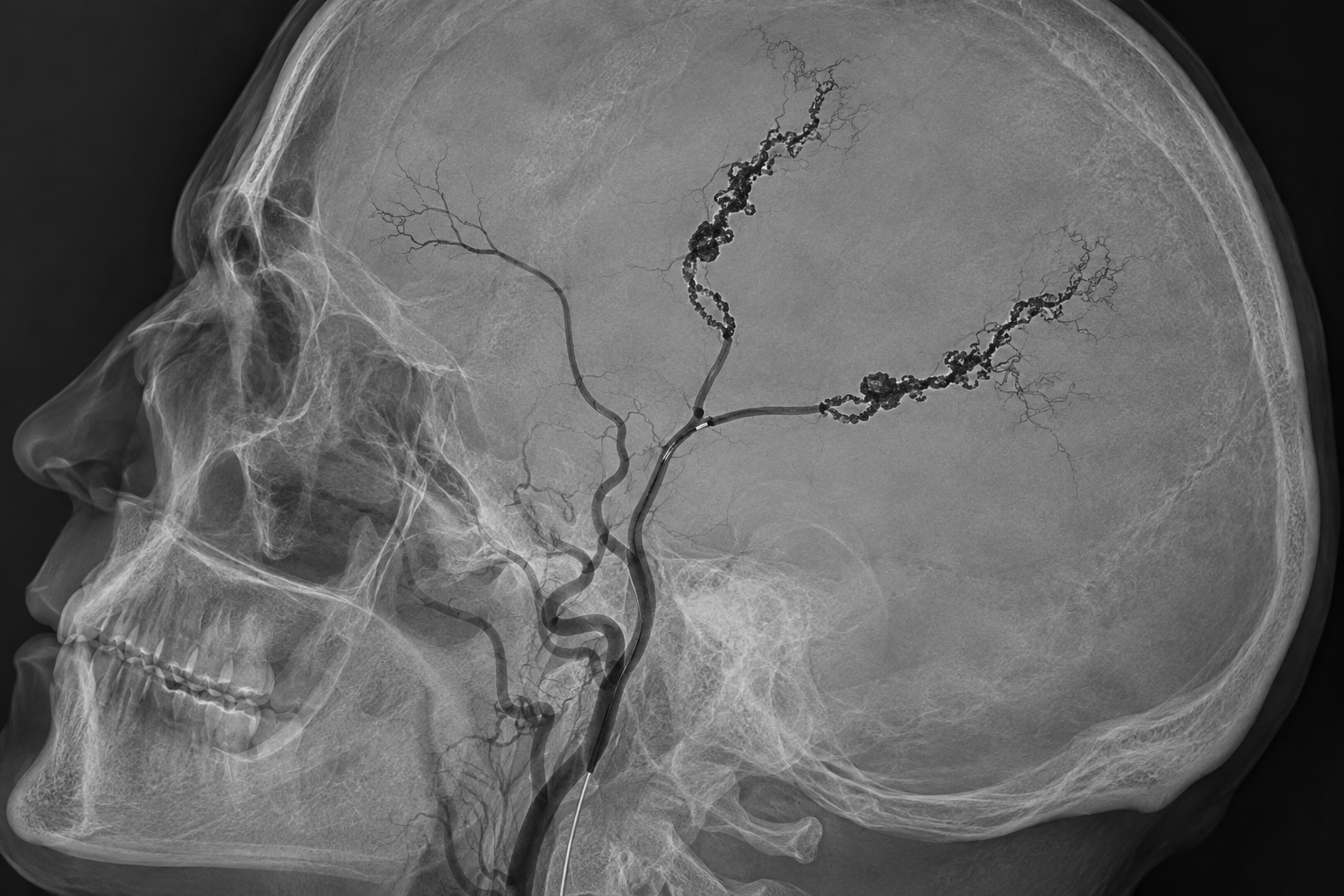
What it is and why it may be advised
A chronic subdural haematoma is a slowly evolving collection of blood and fluid between the brain and its outer covering. Fragile vessels in the surrounding membranes are supplied mainly by the middle meningeal artery (MMA). Embolisation closes selected MMA branches to reduce this abnormal blood supply, helping the collection shrink gradually and lowering the chance of recurrence in appropriately selected patients.
How the strategy is chosen
The neurointerventional and neurosurgical teams review symptoms, neurological stability, CT findings, haematoma size, mass effect, recurrence, frailty and antithrombotic medication. MMA embolisation may be used alongside surgical drainage or, in selected stable patients, as a stand-alone option when surgery is unsuitable. It does not provide immediate decompression and must not delay urgent surgery when there is major mass effect or neurological deterioration.
What happens step by step
- 1
CT confirms the chronic subdural collection, its thickness, density, laterality and any pressure or midline shift. Medicines, kidney function, clotting and the need for urgent surgery are reviewed.
- 2
Through the wrist or groin, a catheter is guided into the external carotid artery. DSA maps the MMA and checks for hazardous connections to the eye, cranial nerves or normal intracranial circulation.
- 3
A microcatheter is advanced into a safe MMA position. The operator selects the branches supplying the haematoma membranes while avoiding dangerous anastomoses.
- 4
A liquid embolic agent such as Onyx, or another suitable material, is delivered slowly under continuous fluoroscopy until the intended abnormal membrane supply is closed.
- 5
Final angiography confirms the treatment result and preserved normal vessels. Neurological and access-site monitoring follow, with interval CT to document gradual reduction of the haematoma.
Options and alternatives
- Observation with repeat clinical assessment and CT for small, stable collections with mild or no symptoms.
- Burr-hole drainage or craniotomy when rapid decompression is required; MMA embolisation may be added to reduce recurrence risk.
- MMA embolisation alone in carefully selected, neurologically stable patients, particularly when surgery or interruption of antithrombotic therapy carries substantial risk.
- Randomised studies including EMBOLISE, STEM and MAGIC-MT support benefit in selected pathways, but results differ by patient group, standard treatment and embolic material.
Procedure-specific risks and complications
- Non-target embolisation through an orbital or cranial-nerve connection can rarely cause visual loss, facial weakness, numbness or other permanent neurological injury.
- Stroke, vessel dissection, perforation, intracranial bleeding or seizure are uncommon but potentially serious complications.
- Groin or wrist bleeding, arterial injury, contrast allergy, kidney injury and sedation or anaesthesia complications.
- Incomplete embolisation, persistent or recurrent haematoma, delayed deterioration or the need for later surgical drainage despite technically successful treatment.
After the procedure
- Symptoms and CT appearance usually improve over weeks rather than immediately; scheduled neurological review and follow-up imaging are important.
- Antiplatelet or anticoagulant interruption and restart are decided individually with the treating teams—never change these medicines without instruction.
- Increasing headache, weakness, confusion, drowsiness, seizure, repeated vomiting or reduced consciousness requires urgent emergency assessment.
04After subarachnoid haemorrhage
Cerebral vasospasm treatment
Open detailed guideClose detailed guide
After subarachnoid haemorrhage
Cerebral vasospasm treatment
Open detailed guideClose detailed guide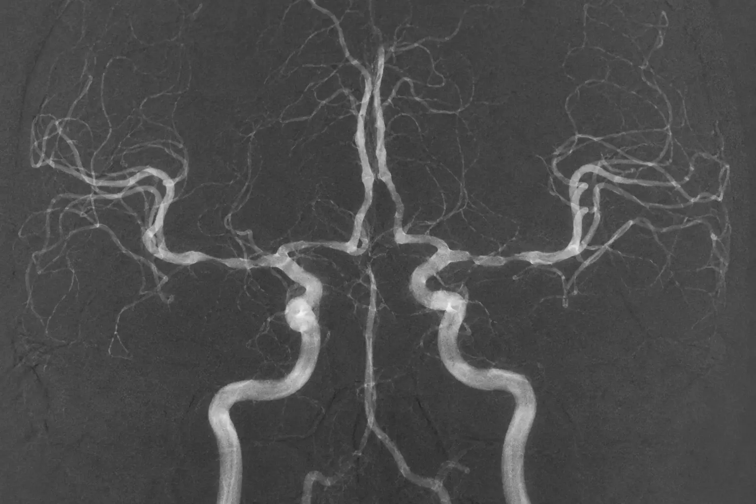
What it is and why it may be advised
After aneurysmal subarachnoid haemorrhage, brain arteries can narrow and reduce blood flow. Delayed cerebral ischaemia is the clinical problem the team aims to prevent and treat; arterial narrowing on a scan is only one part of the assessment.
How the strategy is chosen
Care begins with securing the ruptured aneurysm, oral or enteral nimodipine when appropriate, maintaining normal circulating volume and repeated neurological assessment. CTA, perfusion imaging, transcranial Doppler or DSA may help. Endovascular therapy is reserved for symptomatic or severe vasospasm that does not respond adequately to medical measures.
What happens step by step
- 1
The stroke/neurocritical team identifies new neurological change and excludes other causes such as rebleeding, hydrocephalus, seizure, infection or metabolic disturbance.
- 2
Blood pressure and fluid status are optimised; nimodipine is continued when tolerated and clinically appropriate.
- 3
If threatened brain tissue remains at risk, urgent angiography identifies the narrowed arteries and collateral flow.
- 4
A vasodilator may be infused directly into selected arteries; accessible severe proximal narrowing may also be treated with cautious balloon angioplasty.
- 5
Response is checked angiographically and clinically. Treatment sometimes needs repeating because vasospasm can recur during the risk period.
Options and alternatives
- Close monitoring and guideline-based medical management are the foundation of care.
- Intra-arterial vasodilator treatment can reach distal or multiple narrowed vessels but its effect may be temporary.
- Balloon angioplasty can produce a more durable opening of suitable proximal arteries but cannot safely reach every vessel.
Procedure-specific risks and complications
- Low blood pressure or systemic effects from vasodilator medication, potentially reducing brain perfusion.
- Vessel dissection, perforation or rupture during angioplasty, causing intracranial bleeding.
- Clot formation, embolic stroke or worsening ischaemia despite treatment.
- Recurrent vasospasm and delayed cerebral infarction may occur even after an initially successful procedure.
After the procedure
- Continuous neurocritical or stroke-unit monitoring continues throughout the delayed-ischaemia risk period.
- Serial examination and imaging guide repeat therapy; neurological recovery depends on the original haemorrhage and any secondary infarction.
- After the acute phase, rehabilitation and follow-up address physical, cognitive, emotional and fatigue-related effects.
CT & fluoroscopy-guided spine procedures
Targeted day-care procedures for selected spinal nerve pain and specialist contrast imaging of the spinal canal.
01CT-guided day care
PRT / periradicular injection therapy
Open detailed guideClose detailed guide
CT-guided day care
PRT / periradicular injection therapy
Open detailed guideClose detailed guide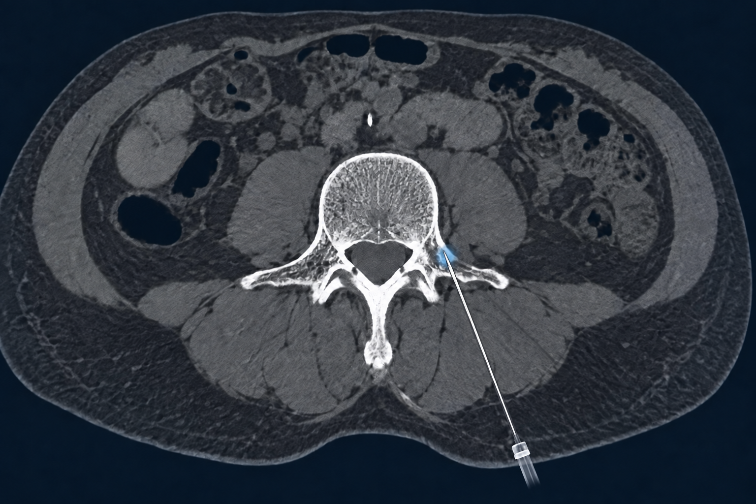
What it is and why it may be advised
PRT places a small amount of local anaesthetic, often with an anti-inflammatory corticosteroid when appropriate, beside a specific spinal nerve root. It may be considered when clinical examination and CT or MRI identify radicular pain that matches an irritated or compressed nerve root. Relief varies and may be temporary; the procedure does not remove a disc prolapse or bony narrowing.
How the strategy is chosen
The target level and side must match the symptoms and imaging. The team reviews weakness or numbness, previous treatment, diabetes, pregnancy, allergy, infection, kidney function when relevant, and medicines that affect clotting. PRT may be therapeutic and may also help establish whether one nerve root is the main pain source. Medication choice and any interruption of antiplatelet or anticoagulant therapy are individual medical decisions.
What happens step by step
- 1
Before arrival, follow the unit’s instructions about eating, drinking, driving and medicines. Bring imaging and an up-to-date medication list; never stop a blood thinner without direct instruction.
- 2
In the day-care unit, identity, symptoms, target level, consent, allergies, pregnancy possibility, observations and any required blood tests are checked.
- 3
You lie on the CT table, usually face down or on your side. The skin is cleaned and local anaesthetic numbs the entry site.
- 4
Short CT images guide a fine needle to the planned position beside the nerve root. A small contrast injection may confirm distribution and help exclude placement in a blood vessel.
- 5
The selected medication is injected slowly, the needle is removed and a small dressing is applied. You are observed until movement, sensation and vital signs are satisfactory.
- 6
You leave with written instructions and an escort if sedation was used or the unit advises it. The clinical effect is reviewed later rather than judged only in the first hours.
Options and alternatives
- Activity modification, suitable analgesia, physiotherapy and treatment of the underlying spinal condition.
- Targeted transforaminal/periradicular injection, interlaminar or caudal epidural injection, depending on the pain pattern and anatomy.
- Specialist spinal or surgical review when there is progressive weakness, cauda-equina concern, structural instability or persistent disabling compression.
- Choosing no injection is reasonable when expected benefit is low or bleeding, infection, allergy or neurological risk is unacceptable.
Procedure-specific risks and complications
- Temporary increase in pain, local bruising, flushing, dizziness, nausea, numbness or weakness; falls are possible until sensation and strength are normal.
- Bleeding or spinal/foraminal haematoma, especially with clotting disorders or antithrombotic medicines; infection or abscess is uncommon.
- Inadvertent vascular, epidural or intrathecal injection; dural puncture can cause a positional headache. Contrast or medication allergy may occur.
- Corticosteroids may temporarily raise blood glucose, disturb sleep or mood, and rarely cause other systemic effects. Benefit may be absent or short-lived.
- Permanent nerve injury, spinal cord injury, infarction, paralysis, seizure or death are very rare but serious complications reported with spinal injections.
After the procedure
- Rest as instructed on the day; avoid driving, hazardous work and strenuous activity until the advised time and until strength and sensation are normal.
- Record pain response over the following days. Local anaesthetic may wear off before any steroid effect begins; follow-up determines the next step.
- Seek urgent help for new or worsening weakness, saddle numbness, loss of bladder/bowel control, severe positional headache, fever, spreading redness, increasing back pain, breathing difficulty or other severe symptoms.
02Fluoroscopy-guided day care
Epidural injection therapy
Open detailed guideClose detailed guide
Fluoroscopy-guided day care
Epidural injection therapy
Open detailed guideClose detailed guide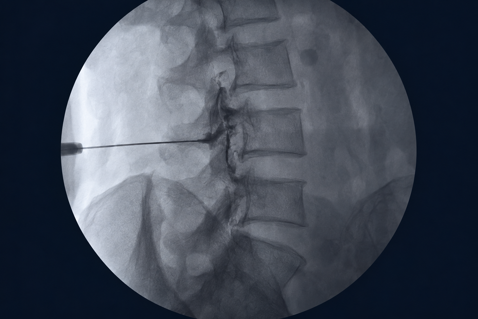
What it is and why it may be advised
An epidural injection delivers local anaesthetic and, when appropriate, corticosteroid into the space outside the dural sac. It may be offered for selected radicular pain from disc disease, spinal stenosis or postoperative inflammation after clinical and imaging review. It aims to reduce inflammation and pain sufficiently to support mobility and rehabilitation; it is not guaranteed and does not correct the underlying anatomy.
How the strategy is chosen
The approach may be interlaminar, caudal or transforaminal according to the affected level, symptoms, previous surgery and safest access. Expected short-term benefit is weighed against diabetes, infection, allergy, pregnancy and bleeding risk. Epidural corticosteroid use requires individual consent, including discussion of rare serious neurological events and the regulatory status of the proposed medicine and route.
What happens step by step
- 1
The day-care team confirms the plan, medication list, clotting safety, allergies, consent and a responsible escort when required.
- 2
You are positioned on the fluoroscopy table. Monitoring is applied, the skin is disinfected and local anaesthetic is given; sedation is used only when planned.
- 3
A fine needle is advanced using live x-ray guidance. Small amounts of contrast confirm the intended epidural pattern and check for vascular or intrathecal spread.
- 4
Medication is injected slowly. Pressure or a brief reproduction of familiar pain can occur and should be reported immediately.
- 5
After needle removal, you are observed for pain, strength, sensation, blood pressure and any medication reaction before discharge.
- 6
Written aftercare explains activity, diabetes monitoring when relevant, medicines, follow-up and symptoms requiring urgent review.
Options and alternatives
- Conservative care with exercise-based rehabilitation, analgesia and management of sleep, work and psychosocial contributors.
- A different image-guided route or a selective nerve-root injection when a more focused diagnostic or therapeutic approach is appropriate.
- Surgical consultation for progressive neurological deficit, cauda-equina symptoms, severe structural compression or persistent disability despite suitable care.
Procedure-specific risks and complications
- Injection-site pain, temporary numbness or weakness, headache, vasovagal reaction, facial flushing, sleep/mood disturbance and transient rise in blood glucose.
- Dural puncture and post-dural-puncture headache; unintended intrathecal or intravascular injection; medication or contrast reaction.
- Bleeding/epidural haematoma, infection, meningitis or epidural abscess are uncommon but may require urgent treatment.
- Rare serious neurological complications include nerve or spinal cord injury, infarction, paralysis, loss of vision, seizure, stroke or death.
- No benefit, incomplete benefit or only temporary relief; repeated steroid exposure has cumulative systemic risks and is not automatically appropriate.
After the procedure
- Arrange transport and follow local driving advice. Resume activity gradually; do not test a numb or weak leg by walking without assistance.
- Patients with diabetes may need closer glucose checks according to their usual clinical plan.
- Urgent assessment is required for progressive weakness, bladder/bowel dysfunction, saddle numbness, severe headache, fever, wound discharge, rapidly increasing pain, breathing difficulty or collapse.
03Diagnostic day care
Myelography and CT myelography
Open detailed guideClose detailed guide
Diagnostic day care
Myelography and CT myelography
Open detailed guideClose detailed guide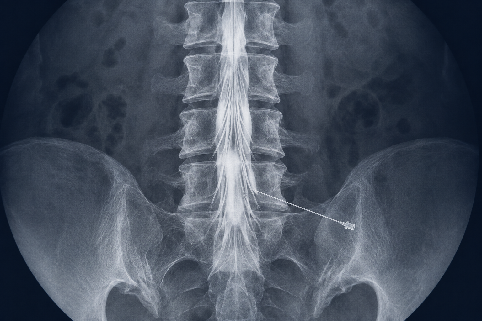
What it is and why it may be advised
Myelography uses fluoroscopy to inject iodinated contrast into the cerebrospinal-fluid space through a lumbar puncture. Fluoroscopic images and usually CT then show the thecal sac, spinal cord and nerve-root sleeves. It is particularly useful when MRI is contraindicated or limited by metal, when symptoms and MRI do not agree, and for selected questions such as dynamic compression or localisation of a cerebrospinal-fluid leak.
How the strategy is chosen
The neuroradiologist confirms that myelography can answer a question that non-invasive imaging cannot answer adequately. Previous imaging, neurological findings, pregnancy possibility, contrast reactions, kidney function when relevant, infection, clotting and medicines are reviewed. The exact technique and positioning vary with the spinal level and clinical question.
What happens step by step
- 1
Follow the department’s instructions about food, fluids and medicines, and arrange transport. Bring previous scans and report contrast reactions, seizures, pregnancy possibility and all medicines.
- 2
In day care, observations, consent and any required laboratory results are checked. You change into a gown and lie on the fluoroscopy table.
- 3
After sterile preparation and local anaesthetic, a fine needle enters the lower lumbar cerebrospinal-fluid space. A sample may be collected only when clinically requested.
- 4
Iodinated contrast is injected slowly. The table and your position may be adjusted while fluoroscopy follows the contrast through the required part of the spinal canal.
- 5
The needle is removed and CT images are commonly obtained soon afterwards. Staff then observe neurological status, puncture site, pain, nausea and headache.
- 6
Discharge instructions cover fluids, activity, medicines, driving and whom to contact. A specialist interprets the images and sends the report to the referring team.
Options and alternatives
- MRI is usually preferred when it can answer the clinical question safely and clearly.
- Non-contrast CT can assess bone and hardware but does not provide the same intrathecal outline of nerves and thecal sac.
- MR myelography or specialised CT/digital-subtraction myelography may be chosen for selected cerebrospinal-fluid leak pathways.
Procedure-specific risks and complications
- Post-lumbar-puncture headache from cerebrospinal-fluid leakage is the common important complication; it may occasionally require an epidural blood patch.
- Temporary back discomfort, nausea, dizziness, vomiting or altered sensation can occur.
- Bleeding, spinal haematoma, infection or meningitis; nerve injury is uncommon.
- Allergic-like contrast reaction and, rarely, seizure or other neurological deterioration. Radiation exposure comes from fluoroscopy and CT.
- A technically limited or non-diagnostic study may require alternative or repeat imaging.
After the procedure
- Remain for the observation period and follow the department’s individual instructions about hydration, rest, medication and returning to normal activity.
- A mild headache may improve with rest and fluids if these are appropriate for you; persistent or severe symptoms need medical advice.
- Seek urgent help for severe or worsening headache, fever, neck stiffness, repeated vomiting, confusion, seizure, new weakness/numbness, bladder/bowel change, wound leakage or breathing difficulty.
In-hospital stroke-unit care
A dedicated stroke unit brings specialist monitoring, prevention and rehabilitation together around the patient.
01Coordinated inpatient pathway
How the dedicated stroke unit works
Open detailed guideClose detailed guide
Coordinated inpatient pathway
How the dedicated stroke unit works
Open detailed guideClose detailed guide
What it is and why it may be advised
A stroke unit is a specialised ward where physicians, stroke-trained nurses and rehabilitation professionals work as one team. Organised stroke-unit care improves outcomes because complications are recognised early and treatment and rehabilitation follow a coordinated plan.
How the strategy is chosen
Care intensity is adapted to stroke type, severity, treatment received and medical stability. Some patients initially need intensive care or high-dependency monitoring before transfer to the stroke unit. The team sets daily priorities and reviews progress with the patient and family.
What happens step by step
- 1
Admission and handover: the emergency, imaging, neurointerventional and anaesthesia teams communicate the diagnosis, treatment result, medicines and immediate risks.
- 2
Close monitoring: repeated neurological examinations, blood pressure, heart rhythm, oxygen, temperature, glucose, fluid balance and puncture-site checks identify early change.
- 3
Safety screening: swallowing is tested before oral intake; mobility, falls, skin integrity, continence, nutrition, communication and cognition are assessed.
- 4
Cause and prevention: vessel and cardiac investigations, blood tests and imaging guide antithrombotic therapy, cholesterol treatment, blood-pressure goals and risk-factor management.
- 5
Early rehabilitation: physiotherapy, occupational therapy, speech and language therapy, dietetics and psychology/neuropsychology begin when safe and set measurable goals.
- 6
Discharge planning: the team reviews function, home support, equipment, medicines, warning signs, driving/work issues and follow-up or inpatient rehabilitation needs.
Options and alternatives
- Stroke-unit care for medically stable patients who need specialist observation, treatment and early rehabilitation.
- Neurocritical/intensive care when ventilation, invasive monitoring, severe brain swelling, major haemorrhage or unstable circulation requires higher support.
- Early supported discharge, inpatient rehabilitation or community rehabilitation according to medical stability, disability, home circumstances and available services.
Procedure-specific risks and complications
- Complications of stroke include brain swelling, haemorrhagic transformation or rebleeding, seizures and neurological deterioration.
- Swallowing problems can cause aspiration and pneumonia; immobility can cause clots, pressure injury, stiffness, falls and loss of strength.
- Heart-rhythm disturbance, infection, dehydration, malnutrition, glucose disturbance, pain, delirium, depression and anxiety may affect recovery.
- Despite optimal care, some injury may be permanent. The team should communicate uncertainty, expected trajectory and changing goals honestly.
After the procedure
- Patients and families participate in goal setting, education and decisions whenever possible.
- A written discharge plan should cover medicines, appointments, rehabilitation, risk-factor targets and what to do if symptoms recur.
- Recovery continues after discharge and may include fatigue, cognitive or emotional difficulties even when physical recovery appears good.
Evidence
Clinical basis and further reading
This educational guide reflects major professional guidance and official patient resources. Recommendations evolve and do not replace an individual consultation.
Medical content prepared for clinician review · 13 September 2026
General education only. Your diagnosis, treatment choice and individual risk must be discussed with your neurovascular team.