
01 · Understanding the condition
What is a CSF leak?
Cerebrospinal fluid (CSF) cushions the brain and spinal cord. When fluid escapes through a spinal dural defect or drains directly into a vein through a CSF–venous fistula, the volume of CSF around the brain can fall. The resulting syndrome is called spontaneous intracranial hypotension (SIH).
Despite the name, lumbar-puncture opening pressure can be normal. Diagnosis therefore depends on the clinical pattern and the complete imaging pathway—not on one pressure measurement alone.
02 · Recognition
Symptoms are broader than positional headache
The classic symptom is an orthostatic headache: pain develops or worsens after becoming upright and improves when lying flat. The positional pattern may become less obvious in long-standing disease.
- Neck pain or stiffness, nausea and sensitivity to light
- Tinnitus, muffled hearing, dizziness or imbalance
- Blurred or double vision
- Cognitive slowing, fatigue or a feeling of pressure
- Rarely, marked drowsiness, movement disorder or other severe neurological deterioration
03 · Leak mechanisms
Dural tears, diverticula and CSF–venous fistulas
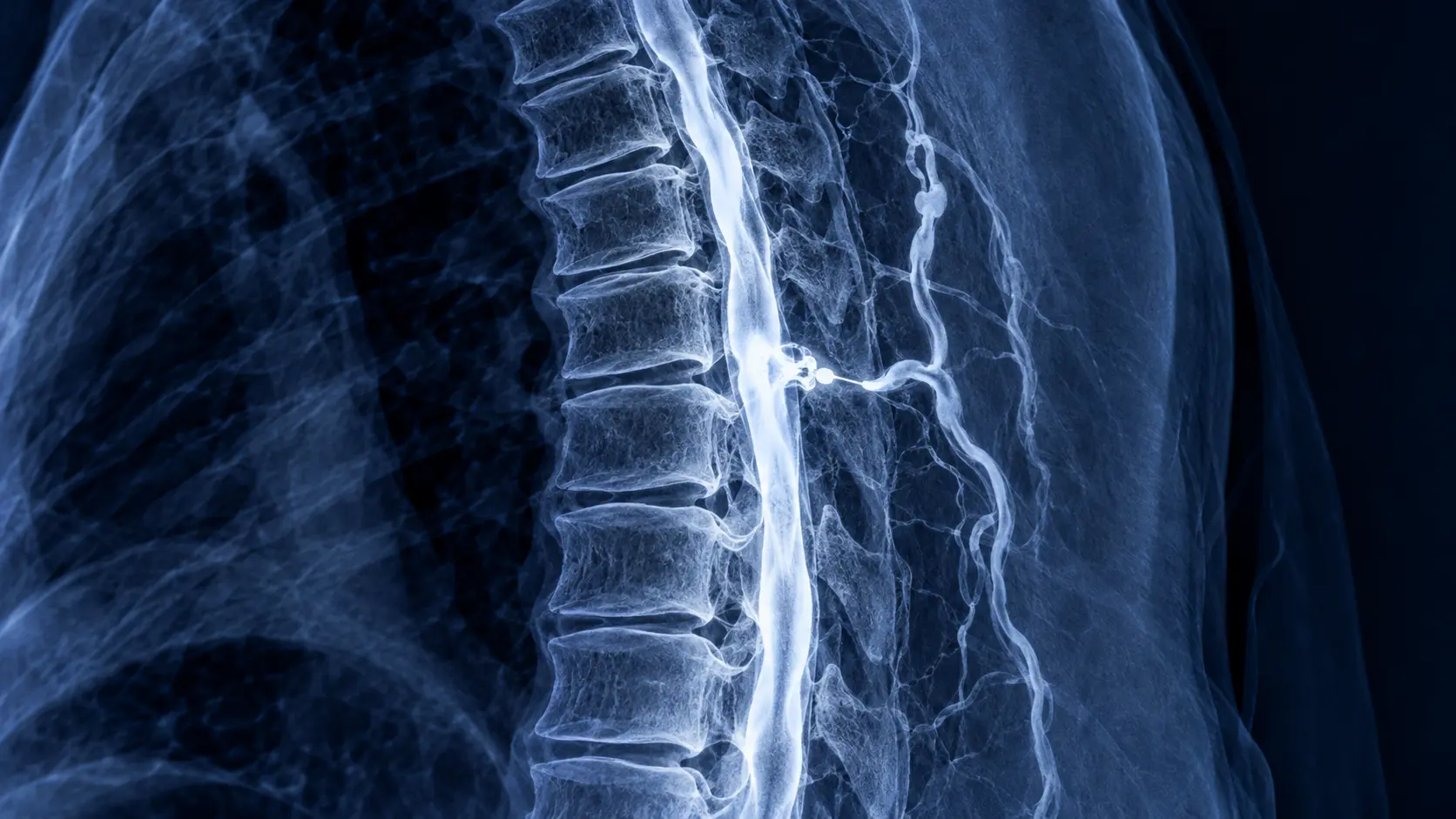
A ventral dural tear may be caused by a small calcified disc or bony spur. A meningeal diverticulum can leak into the surrounding tissues. In a CSF–venous fistula, fluid passes directly from a nerve-root sleeve into an adjacent vein, often without a visible extradural fluid collection.
Previous lumbar puncture, spinal or epidural procedure, surgery and trauma can also cause a leak. Connective-tissue vulnerability may be relevant in selected patients, but many people have no obvious systemic disorder.
04 · Neuroimaging pathway
From brain MRI to specialised myelography
The usual first-line imaging is contrast-enhanced brain MRI together with whole-spine MRI. Brain findings may include smooth diffuse pachymeningeal enhancement, venous engorgement, pituitary enlargement, subdural collections and downward displacement or “sagging” of the brain. MRI can be normal, so a normal scan does not completely exclude SIH.
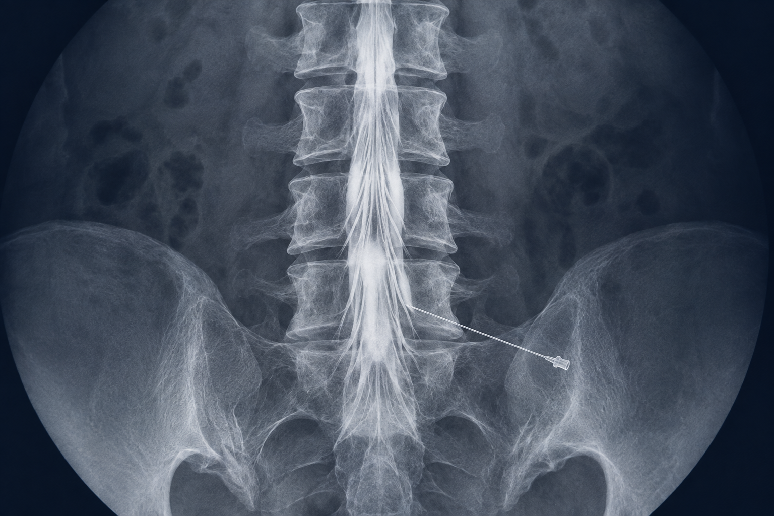
Spine MRI looks for extradural CSF and helps select the next test. When precise localisation is required, dynamic CT myelography or digital-subtraction myelography is tailored to the suspected leak type. Lateral-decubitus techniques are particularly important for detecting CSF–venous fistulas. MR myelography is useful in selected pathways.
05 · Treatment
Treatment is matched to the leak mechanism
Initial management may include short-term activity modification, hydration and caffeine when appropriate, but prolonged conservative treatment should not delay specialist care in a disabling case. A high-volume non-targeted epidural blood patch is a common first-line procedure; a targeted patch or fibrin sealant may be chosen once the leak is localised.
Persistent structural tears or diverticular leaks may require microsurgical repair. For a suitable CSF–venous fistula, intervention may be surgical or minimally invasive transvenous embolisation. During embolisation, a microcatheter is navigated through the venous system and liquid embolic material closes the abnormal fistulous connection. Evidence is promising but continues to evolve, and patient selection should be multidisciplinary.
06 · After treatment
Recovery and rebound pressure
Symptoms do not always resolve immediately, and follow-up may include clinical assessment and repeat imaging. After successful closure, some patients develop rebound intracranial hypertension: the previous upright headache changes to a headache that is worse lying down or at night, sometimes with nausea or visual symptoms.
New visual disturbance, severe worsening headache, repeated vomiting or neurological symptoms after treatment should be reported promptly. The treating team distinguishes rebound pressure from persistent leak or another cause and adjusts treatment accordingly.
07 · Evidence