
01 · Overview
What is IIH?
Idiopathic intracranial hypertension (IIH) is a disorder in which pressure inside the skull is raised, while brain imaging and cerebrospinal fluid testing do not reveal a tumour, hydrocephalus, infection or another direct cause. The older terms ‘pseudotumour cerebri’ and ‘benign intracranial hypertension’ are now used less often: IIH is not a tumour, but it is not always benign because untreated optic-nerve swelling can cause permanent sight loss.
Pressure-related symptoms may fluctuate, and headache severity does not reliably show how much the optic nerves are at risk. Vision therefore needs to be assessed separately and followed objectively.
02 · Recognition
Signs and symptoms
Headache is common and may resemble migraine. Some people describe daily pressure, pain that is worse on waking, bending, coughing or straining, neck discomfort, nausea or light sensitivity.
- Brief dimming or greying of vision, often after standing or bending
- Blurred vision, loss of peripheral vision or reduced colour/contrast perception
- Double vision, sometimes from a sixth cranial nerve palsy
- Pulse-synchronous ‘whooshing’ tinnitus
- Papilloedema—swelling of both optic discs seen during an eye examination
03 · Risk profile
Who is more often affected?
IIH occurs most often in women of reproductive age who are living with obesity, particularly after recent weight gain. However, it can also occur in men, children, adolescents and people with a normal body weight.
Obstructive sleep apnoea, anaemia and polycystic ovary syndrome are commonly encountered in patients with IIH. These associations do not mean that each condition directly causes IIH. When the clinical profile is atypical, the medical team looks especially carefully for a secondary cause of raised intracranial pressure.
04 · Medicines & hormones
Can medicines or hormones play a role?
Some medicines can produce a syndrome that resembles IIH and should be reviewed with the prescribing clinician. The strongest recognised associations include tetracycline antibiotics such as doxycycline or minocycline, vitamin A derivatives such as isotretinoin, recombinant growth hormone and lithium. Raised pressure has also been reported after withdrawal of long-term corticosteroids.
Metabolic and hormonal pathways—including androgen signalling and adipose-tissue biology—are active areas of research. PCOS is more common among women with IIH, but its precise causal role remains uncertain. Current evidence does not show that hormonal contraception increases the prevalence of IIH; it should not be stopped automatically without individual medical advice.
05 · Diagnostic pathway
How the diagnosis is established
Diagnosis is multidisciplinary. It usually combines a neurological and neuro-ophthalmological assessment, dilated fundus examination, formal visual-field testing and optical coherence tomography (OCT). Blood tests may look for anaemia and other secondary factors.
Imaging is performed before lumbar puncture. When it is safe and clinically indicated, lumbar puncture measures the opening pressure with the patient correctly positioned and checks that cerebrospinal fluid composition is normal. A pressure value is interpreted together with symptoms, eye findings and imaging—not in isolation.
06 · Neuroimaging
The role of MRI and MR venography
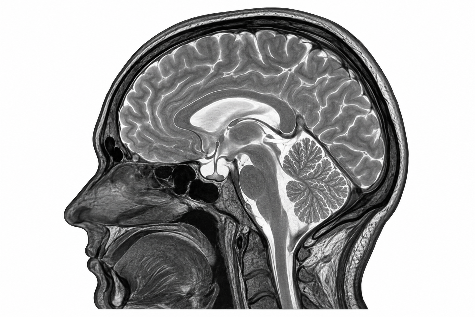
Brain MRI helps exclude a mass, hydrocephalus, inflammation and other structural causes. Supportive signs of raised pressure can include a partial or pseudo-empty sella, flattening of the posterior globes, distension or tortuosity of the optic-nerve sheaths and protrusion of the optic nerve heads.
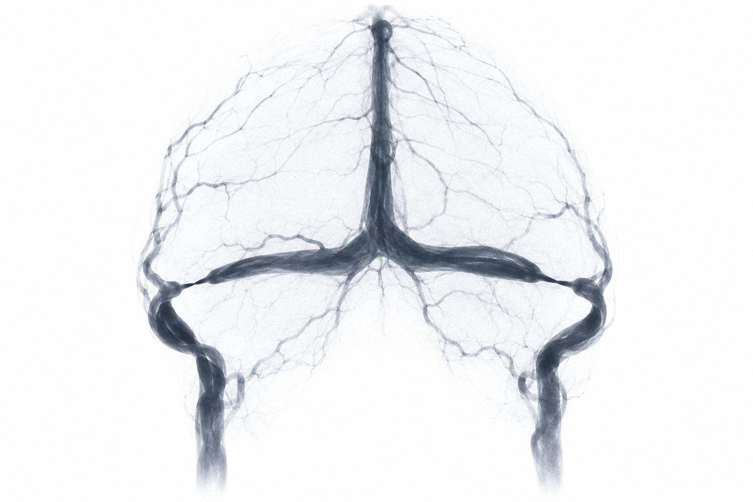
MR venography (MRV) is essential for excluding cerebral venous sinus thrombosis, which requires different treatment. It also assesses narrowing of the transverse sinuses. Transverse-sinus stenosis is common in IIH and may both contribute to and result from raised pressure; its presence alone does not prove the diagnosis.
07 · Advanced assessment
When are catheter venography and DSA used?
Catheter cerebral venography/venous-phase DSA is not the routine first test. It may be considered in carefully selected patients with persistent symptoms, threatened vision or significant transverse-sinus narrowing when venous sinus stenting is being evaluated.
During the same procedure, pressure measurements can be taken on both sides of a stenosis. The anatomy, pressure gradient, symptoms, eye findings and response to medical treatment are reviewed together by a multidisciplinary team before any intervention is proposed.
08 · Treatment
Treatment is tailored to the individual
The priorities are to protect vision, reduce pressure and manage headache disability. Where appropriate, sustainable weight reduction is the only established disease-modifying strategy. Acetazolamide is commonly used to reduce cerebrospinal fluid production; topiramate may be considered in selected patients, especially when migraine-like headache is prominent.
Rapidly declining vision is an emergency. Optic-nerve sheath fenestration or cerebrospinal-fluid diversion may be required. Venous sinus stenting is an option for selected patients with medically refractory disease, relevant venous stenosis and a significant pressure gradient—not a universal first-line treatment.
09 · Evidence