
01 · Overview
What is intracranial atherosclerotic stenosis?
Intracranial atherosclerotic stenosis (ICAS), also called intracranial atherosclerotic disease, is plaque-related narrowing of a major artery within the skull. Common sites include the intracranial internal carotid, middle cerebral, vertebral and basilar arteries.
A stenosis is described as symptomatic when it is considered responsible for a recent transient ischaemic attack (TIA) or ischaemic stroke in the territory it supplies. A narrowing found incidentally without a matching event is managed differently; the percentage alone does not define the individual risk.
02 · Recognition
Symptoms are a stroke warning
Symptoms depend on the affected artery and may be brief or persistent. Use BE FAST: sudden Balance difficulty, Eye or visual loss, Facial droop, Arm or leg weakness, Speech difficulty—Time to call emergency services.
- Sudden weakness or numbness on one side
- New difficulty speaking, understanding or seeing
- Sudden imbalance, double vision, vertigo with other neurological signs or loss of coordination
- A TIA that resolves still requires urgent assessment
03 · Mechanism & risk
How can the narrowing cause stroke?
Plaque can release clot or debris into smaller downstream arteries, block tiny perforating branches, or reduce flow when collateral circulation cannot compensate. More than one mechanism may coexist.
High blood pressure, diabetes, raised LDL cholesterol, smoking, kidney disease, physical inactivity and increasing age are important risk factors. ICAS can affect anyone, although its frequency varies between populations. The location, severity, recent symptoms, brain infarct pattern and collateral circulation are assessed together.
04 · Systemic vascular health
The connection with coronary arteries and the aorta
ICAD is often one expression of a wider, multi-territory atherosclerotic process. In a prospective stroke registry, patients with significant ICAD more often had coronary atherosclerosis and aortic-arch plaque than those without significant ICAD. When coronary artery disease coexisted with ICAD, subsequent major vascular events were more frequent. This does not mean that every patient has obstructive coronary disease, but it supports a global cardiovascular assessment rather than treating the brain artery in isolation.
Depending on symptoms and overall risk, the treating team may assess blood pressure, lipids, diabetes, kidney function, ECG and cardiac history, and may request cardiology review, coronary testing or aortic imaging. New chest pressure, exertional breathlessness, unexplained sweating or pain radiating to the arm or jaw needs prompt medical assessment.
Aortic aneurysms share several risk factors with ICAD—especially age, smoking, hypertension and systemic vascular disease—but aneurysm formation has distinct degenerative, inflammatory and genetic mechanisms and is not simply caused by an intracranial stenosis. ICAD alone is therefore not an indication for routine CT screening of the entire aorta. Abdominal aortic ultrasound or thoracic aortic imaging is considered according to established criteria such as age, sex, smoking exposure, family history, known aortic disease, connective-tissue or bicuspid-aortic-valve disease, examination findings and symptoms.
05 · Diagnosis
A symptom leads to a tailored imaging pathway
Non-contrast CT or brain MRI identifies haemorrhage and established or recent ischaemic injury. CT angiography (CTA) or MR angiography (MRA) then evaluates the arterial lumen; perfusion CT, perfusion MRI or arterial-spin labelling may be added when the haemodynamic effect is uncertain.
High-resolution vessel-wall MRI can show eccentric plaque and may help distinguish atherosclerosis from dissection, vasculitis, reversible cerebral vasoconstriction, moyamoya-pattern arteriopathy and other causes of narrowing. Findings must always be interpreted with the clinical picture and laboratory assessment.
06 · Angiographic screening
Why assess the whole head-and-neck arterial system?
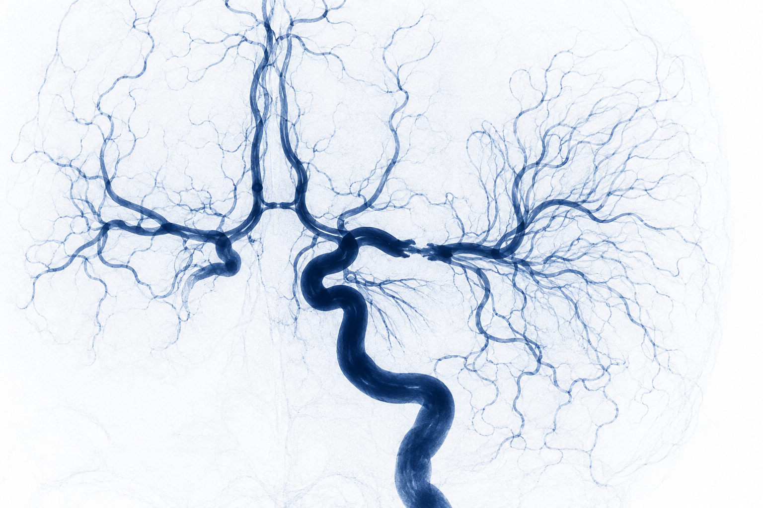
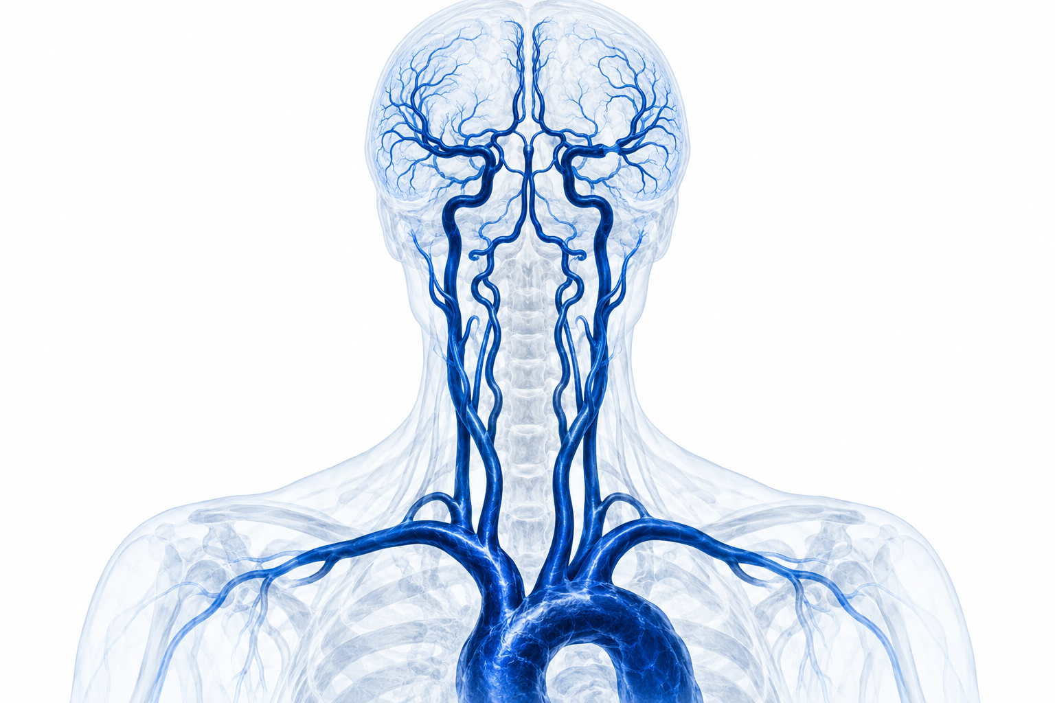
After an intracranial stenosis is identified, a comprehensive non-invasive angiographic survey—often CTA or MRA from the aortic arch to the vertex—can check the extracranial carotid and vertebral arteries, the remaining intracranial segments, tandem or multifocal stenoses and the routes of collateral blood flow. It can also help exclude alternative or coexisting pan-arterial disease rather than assuming that every narrowing is atherosclerosis.
Catheter digital-subtraction angiography (DSA) provides the highest spatial and temporal detail and can assess collateral flow and treatment anatomy. Because it is invasive, it is not routine screening for every patient. It is reserved for selected cases when non-invasive tests are inconclusive or discordant, symptoms recur despite treatment, or an endovascular procedure is being considered.
07 · Prevention & follow-up
Medical prevention comes first
For symptomatic ICAS, first-line care is aggressive, clinician-supervised medical management: appropriate antiplatelet therapy, high-intensity lipid lowering, careful blood-pressure and diabetes control, smoking cessation, physical activity and support with adherence. In selected patients with severe recent symptomatic stenosis, a short course of dual antiplatelet therapy may be prescribed; it should never be started, extended or stopped without the treating team.
Routine intracranial stenting is not the initial treatment for most patients. Angioplasty or stenting may be discussed only in carefully selected patients with recurrent territory-specific ischaemia despite genuinely optimised treatment, favourable anatomy and review by an experienced multidisciplinary neurovascular team. Follow-up combines symptoms, neurological review and selected CTA, MRA or transcranial Doppler; the interval is individual rather than a universal schedule.
08 · Evidence