
01 · Overview
What is multiple sclerosis?
Multiple sclerosis (MS) is a chronic immune-mediated condition of the central nervous system. Inflammation can damage myelin—the insulating layer around nerve fibres—and sometimes the nerve fibres themselves. The effects differ greatly between people and can change over time.
MS is not contagious and is not caused by one lifestyle choice. Genetics, immune biology and environmental exposures all contribute, but no single factor explains every case.
02 · Recognition
Symptoms and warning patterns
Symptoms depend on which part of the central nervous system is affected. A relapse usually means a new or clearly worse neurological symptom lasting more than 24 hours, without fever or another infection, after a period of stability.
- Blurred or painful vision in one eye, colour desaturation or double vision
- Numbness, tingling, weakness, stiffness or imbalance
- Fatigue, bladder symptoms, vertigo or coordination difficulty
- Problems with concentration, memory, mood or sexual function
03 · Clinical course
CIS, relapsing and progressive forms
A clinically isolated syndrome (CIS) is a first neurological episode suggestive of demyelination. Relapsing-remitting MS causes attacks followed by partial or full recovery. Some people later develop secondary progressive MS, with gradual disability independent of clear relapses. Primary progressive MS shows gradual progression from onset.
A radiologically isolated syndrome (RIS) describes MRI lesions typical of demyelination in a person without matching symptoms. It is not automatically MS and requires specialist review and follow-up.
04 · Diagnosis
How the diagnosis is made
MS remains a diagnosis of exclusion. The neurologist combines the history and examination with MRI, and when needed cerebrospinal fluid, optical coherence tomography or evoked potentials. The goal is to demonstrate disease in different central nervous system locations and at different times while ruling out vascular, infectious, metabolic, genetic and other inflammatory mimics.
The 2024 McDonald criteria expand the role of the optic nerve and allow carefully defined supportive markers, including cerebrospinal-fluid oligoclonal bands or kappa free light chains, the central vein sign and paramagnetic rim lesions. These features must be interpreted by an experienced team—not used as stand-alone screening tests.
05 · Neuroimaging
Brain and spinal MRI
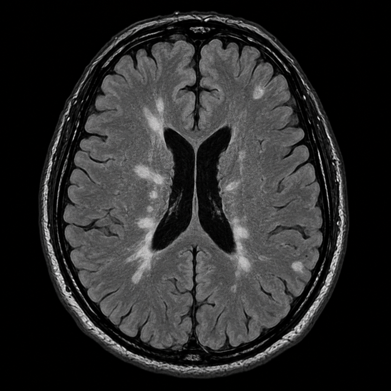
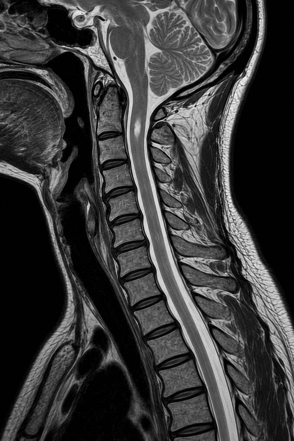
MRI of the brain and, when indicated, cervical and thoracic spinal cord is the key imaging test. Typical plaques may be periventricular and ovoid, juxtacortical or cortical, infratentorial, within the optic pathway or within the spinal cord. Gadolinium enhancement can identify recent inflammatory activity; comparison with prior scans shows change over time.
High-quality protocols and consistent follow-up are important. Small nonspecific white-matter spots are common with migraine, age and vascular risk factors and should not be labelled MS solely because they are bright on FLAIR.
06 · Treatment
Treatment and follow-up
Disease-modifying therapies reduce future inflammatory activity and relapses. Choice depends on disease activity, safety profile, pregnancy plans, other conditions and patient preference. A significant relapse may be treated with corticosteroids after infection has been considered.
Rehabilitation, exercise, sleep, vaccination, smoking cessation and targeted treatment of fatigue, pain, spasticity, mood, bladder or cognitive symptoms are part of comprehensive care. Neurological review and planned MRI monitoring assess response and safety.
07 · Evidence