
01 · Overview
What are pituitary adenomas?
Pituitary adenomas are tumours arising from hormone-producing cells of the anterior pituitary gland. The modern term pituitary neuroendocrine tumour (PitNET) is also used. Most are non-metastatic and can be controlled with observation, medication, surgery or a combination tailored to the tumour and the patient.
Size describes anatomy, not behaviour: microadenomas are under 10 mm, while macroadenomas are 10 mm or larger. A small functioning tumour can cause major hormonal symptoms, whereas a larger non-functioning tumour may first become apparent through visual or pituitary-compression symptoms.
02 · Hormonal activity
Functioning and non-functioning tumours
A functioning tumour releases excess hormone. Prolactinomas may cause irregular or absent periods, milk production, infertility, reduced libido or erectile dysfunction. Growth-hormone excess can cause acromegaly, with enlargement of hands or feet, facial change, sweating, joint pain, sleep apnoea, diabetes or hypertension. ACTH-producing tumours cause Cushing disease, while TSH-producing tumours are uncommon and can cause hyperthyroidism.
A non-functioning tumour does not produce a clinically active hormone, but a macroadenoma can compress the normal gland and reduce one or more pituitary hormones. Symptoms may include fatigue, weakness, menstrual or sexual dysfunction, low blood pressure, cold intolerance or loss of body hair.
03 · Pressure effects
Headache, vision and nearby structures
A macroadenoma may extend above the sella toward the optic chiasm. This can reduce peripheral vision—classically affecting the outer halves of both visual fields—although patients may not notice early change. Extension toward the cavernous sinus can affect eye movements and cause double vision.
Headache is common but does not reliably indicate tumour size or growth. Formal visual-field testing is recommended when a lesion touches or compresses the optic nerves or chiasm, or when visual symptoms are present.
04 · Diagnostic pathway
Hormone tests and specialist assessment
Assessment usually includes a clinical history, examination and blood tests for hormone excess and hormone deficiency. Depending on the presentation, these may include prolactin, IGF-1, morning cortisol with ACTH, thyroid function and the gonadal axis. Some diagnoses need dynamic suppression or stimulation tests rather than a single blood result.
The imaging, endocrine results, symptoms and visual findings are reviewed together. Incidental lesions are common, and not every small pituitary finding is an adenoma or needs treatment.
05 · Neuroimaging
Why a dedicated pituitary MRI matters
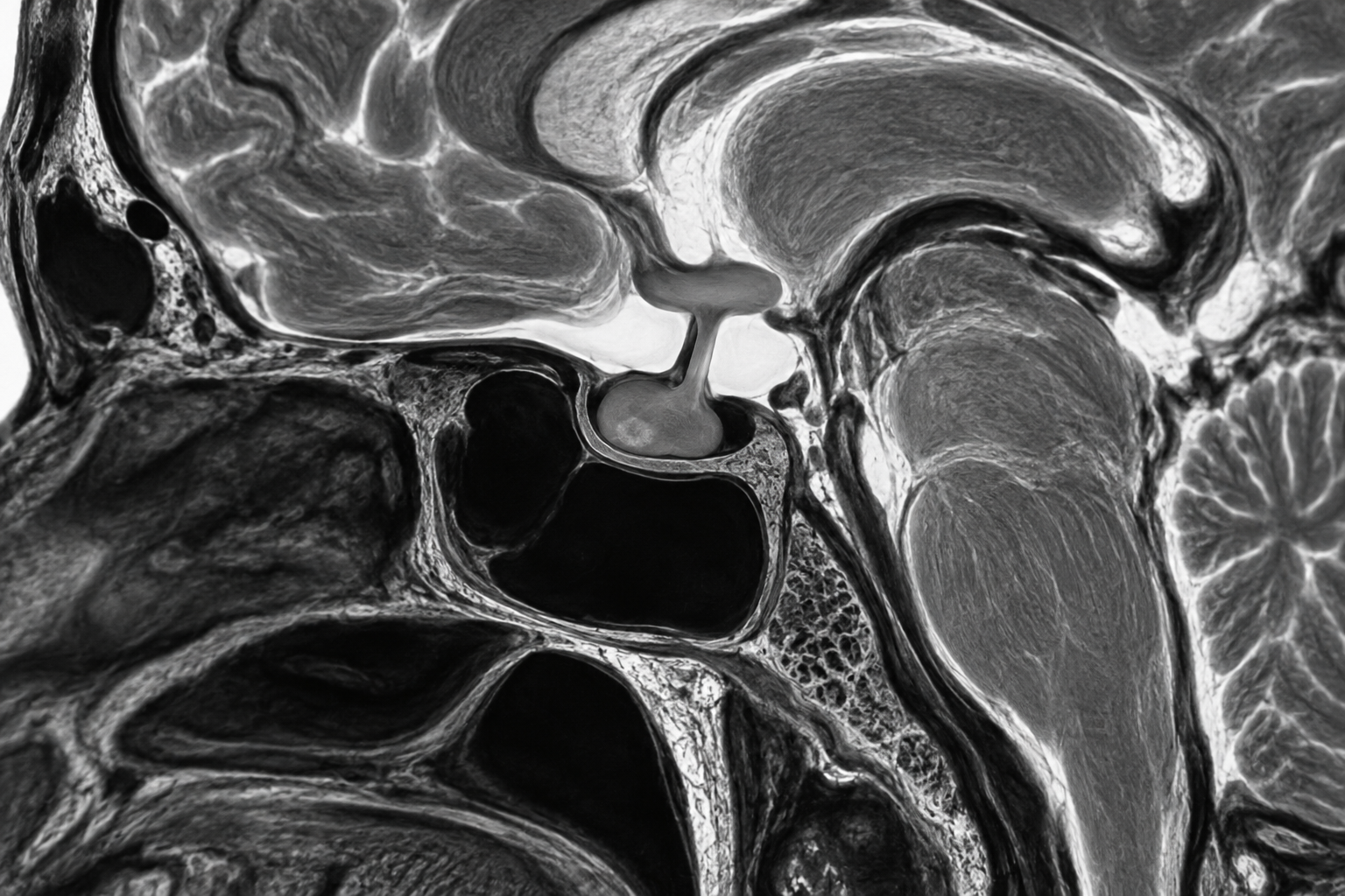
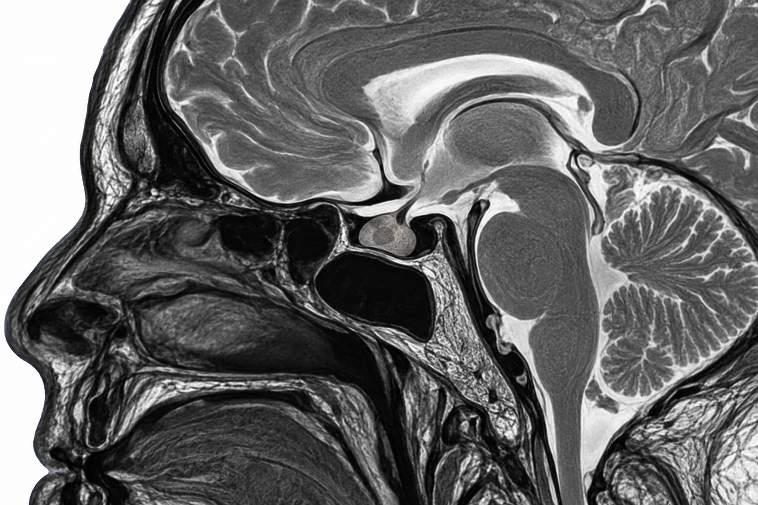
A dedicated sellar MRI uses a small field of view and thin sections, usually in sagittal and coronal planes. T2-weighted images show the gland, stalk, optic chiasm, sphenoid sinus and lesion texture. T1-weighted images before and after gadolinium—sometimes with dynamic early post-contrast imaging—are particularly important for detecting small microadenomas.
For macroadenomas, MRI maps suprasellar extension, optic-chiasm compression, cavernous-sinus relationship, carotid-artery encasement, haemorrhage and the amount of normal gland that remains. A routine brain MRI may detect a large lesion but is less detailed than a dedicated sellar protocol.
06 · Treatment
Observation, medicine, surgery and radiotherapy
Selected small non-functioning lesions can be monitored with clinical, hormonal and MRI follow-up. Most prolactinomas respond to a dopamine agonist such as cabergoline, often reducing both prolactin and tumour size. Other functioning tumours and lesions causing visual or other compressive symptoms commonly require transsphenoidal surgery through the nose.
Specialist medicines may be used before or after surgery for acromegaly, Cushing disease or TSH-secreting tumours. Radiotherapy is reserved for selected residual, recurrent or difficult-to-control disease. The plan is individualised by endocrinology, neurosurgery, neuroradiology, ophthalmology and, when needed, radiation oncology.
07 · Follow-up
Monitoring is based on risk, not size alone
Follow-up intervals depend on tumour type, size, proximity to the optic chiasm, growth, hormonal activity, symptoms, age and treatment. Reviews may include repeat MRI, hormone testing and visual fields. Pregnancy or plans for pregnancy should be discussed early because monitoring and medication choices can change.
A new or worsening symptom should prompt reassessment rather than waiting for the next planned scan.
08 · Evidence