01 · Overview
What is tinnitus?
Tinnitus is the perception of sound when no corresponding external sound is present. It may be heard in one ear, both ears or inside the head, and may be constant or intermittent. For many people it accompanies hearing loss or noise exposure; for others it is linked to a treatable ear, neurological or vascular condition.
Tinnitus is a symptom, not a diagnosis. The first task is to understand the character of the sound, when it began, whether it is one-sided and whether it follows the heartbeat. These details guide examination and determine whether imaging is useful.
02 · Recognition
Pulsatile and non-pulsatile tinnitus are different pathways
Non-pulsatile tinnitus is usually described as ringing, buzzing or hissing. When it is bilateral, symmetrical and associated with age-related or noise-related hearing loss, imaging is often unnecessary unless other concerning signs are present.
Pulsatile tinnitus is rhythmic and usually synchronous with the pulse. It may sound like whooshing, beating or flowing water. Because it can arise from altered arterial or venous flow, a vascular shunt or a vascular tumour, persistent pulsatile tinnitus should be medically assessed and generally imaged.
03 · Possible causes
Common, structural and vascular explanations
Non-pulsatile tinnitus commonly accompanies sensorineural hearing loss, noise exposure, ear disease or medicines that can affect hearing. Unilateral tinnitus or asymmetric hearing loss raises the question of a retrocochlear cause, including a vestibular schwannoma, although most scans do not show a tumour.
Pulsatile tinnitus may relate to venous sinus stenosis, idiopathic intracranial hypertension, sigmoid sinus wall abnormalities, a high jugular bulb, carotid atherosclerosis or dissection, aneurysm, arteriovenous malformation, dural arteriovenous fistula or a vascular middle-ear/skull-base tumour such as a paraganglioma. CT can also identify bony causes such as superior semicircular canal dehiscence.
04 · Warning signs
When assessment should be urgent
Seek urgent medical care when tinnitus appears with sudden hearing loss, new facial weakness, severe vertigo, focal neurological symptoms, confusion, collapse or a sudden severe headache. Tinnitus after significant head or neck trauma also needs prompt assessment.
A new persistent pulse-synchronous sound, particularly when one-sided or accompanied by headache, visual disturbance, neck pain or a sound audible to an examiner, should not be dismissed. Some vascular causes are benign, while others require timely treatment to reduce neurological risk.
- Sudden loss or rapid deterioration of hearing
- Weakness, numbness, speech or balance disturbance
- Severe new headache, neck pain or symptoms after trauma
- Pulse-synchronous sound with visual symptoms or neurological change
- Ear discharge, severe ear pain, fever or a visible mass behind the eardrum
05 · First assessment
History, ear examination and hearing tests come first
The pathway usually begins with history, otoscopy and formal audiometry. The clinician asks about laterality, pulse synchrony, hearing change, dizziness, headache, medicines, noise exposure, trauma and cardiovascular risk. Examination may include the ears, cranial nerves, neck vessels and listening for a bruit.
This first assessment selects the imaging protocol. A single generic brain scan is not the best test for every type of tinnitus, and a normal routine MRI does not exclude every vascular or temporal-bone cause.
06 · Tailored neuroimaging
MRI, CT, angiography and venography each answer a different question
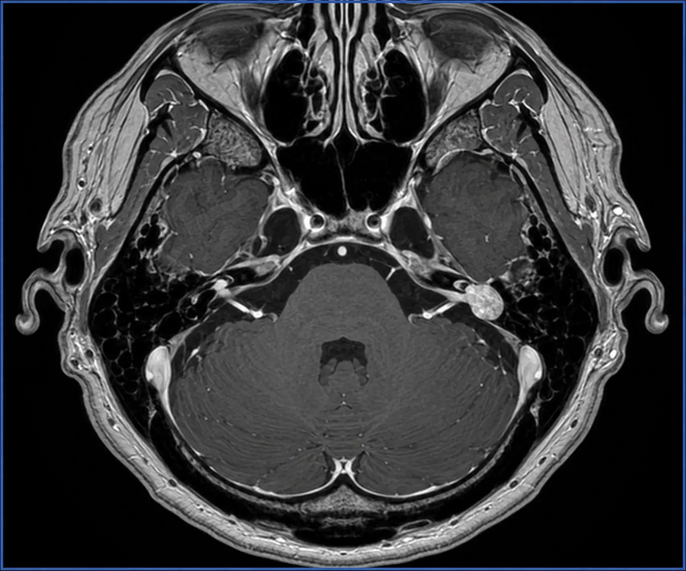
For unilateral non-pulsatile tinnitus or asymmetric sensorineural hearing loss, high-resolution MRI of the brain and internal auditory canals—usually before and after contrast—is the preferred examination for the cochlear nerves, internal auditory canals and cerebellopontine angles.
For pulsatile tinnitus, CTA/CTV or MRI with MRA/MRV can evaluate the arteries, veins, dural sinuses, skull base and soft tissues. Thin-section temporal-bone CT is particularly valuable when otoscopy suggests a retrotympanic lesion or when a bony or sinus-wall abnormality is suspected. The protocol should cover the relevant intracranial and cervical vessels rather than a small region alone.
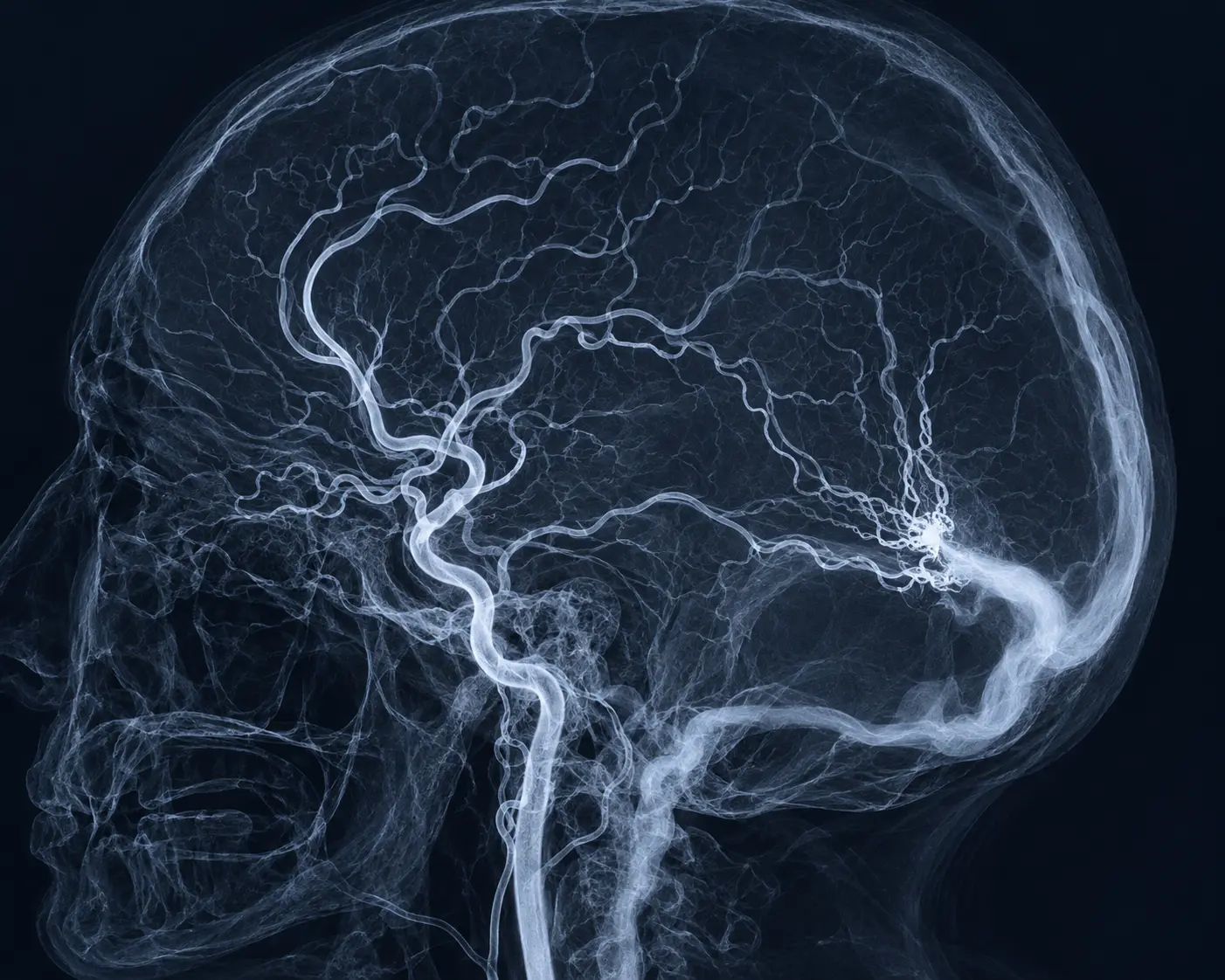
Digital subtraction angiography (DSA) is invasive and is not the routine first test. It is reserved for selected patients when non-invasive imaging is inconclusive but clinical concern for a vascular shunt remains, or when precise vascular anatomy is required to plan treatment. Dynamic catheter angiography remains the reference standard for defining a dural arteriovenous fistula.
07 · Treatment
Treatment targets the cause—and the impact on daily life
When a cause is identified, treatment is specific: hearing rehabilitation for hearing loss; medical and lifestyle management for idiopathic intracranial hypertension; surgery for selected tumours or structural abnormalities; and endovascular or surgical treatment for appropriate vascular lesions. A dural arteriovenous fistula may be treated by embolisation after detailed angiographic assessment.
When no dangerous or reversible cause is found, tinnitus can still be treated. Hearing aids, sound therapy, tinnitus-focused education and cognitive behavioural therapy can reduce distress and improve sleep and quality of life. The goal is not to imply that the symptom is imaginary, but to reduce the brain's threat response and the symptom's functional burden.
08 · Evidence