
01 · World Spine Day
Spine Health for Life
World Spine Day is marked on 16 October. Its 2026 theme, ‘Spine Health for Life’, highlights prevention, physical activity, safe movement and access to evidence-based care throughout life.
Low back pain is extremely common and is the leading cause of disability worldwide. Most episodes are non-specific: a serious structural cause is not identified, and recovery is supported by reassurance, continued activity as tolerated and rehabilitation.
02 · Recognition
When back pain needs urgent assessment
A clinician looks beyond the pain score for signs of nerve-root, spinal-cord or cauda-equina dysfunction and for clues to fracture, infection, inflammation or tumour.
- New or progressive leg or arm weakness, including foot drop
- New bladder retention or incontinence, loss of bowel control or numbness around the saddle area
- Marked gait imbalance, loss of hand dexterity or symptoms in both legs
- Fever, immunosuppression, recent serious infection or intravenous drug use
- Known cancer, unexplained weight loss or persistent night pain
- Significant trauma, osteoporosis or long-term corticosteroid use
03 · Neurological deficit
Foot drop is more than pain
Foot drop means difficulty lifting the front of the foot. It may arise from compression of a lumbar nerve root—often L4 or L5—but can also result from a peripheral nerve lesion or a problem higher in the nervous system.
New foot drop should be examined promptly to document muscle strength, reflexes and sensation and to identify the likely level. Rapidly worsening weakness, bilateral symptoms or associated bladder, bowel or saddle symptoms increase the urgency.
04 · Tailored imaging
CT and MRI answer different questions
CT is fast and excellent for fractures, alignment and detailed bone anatomy. It is often the first choice after significant trauma or when MRI cannot be performed.
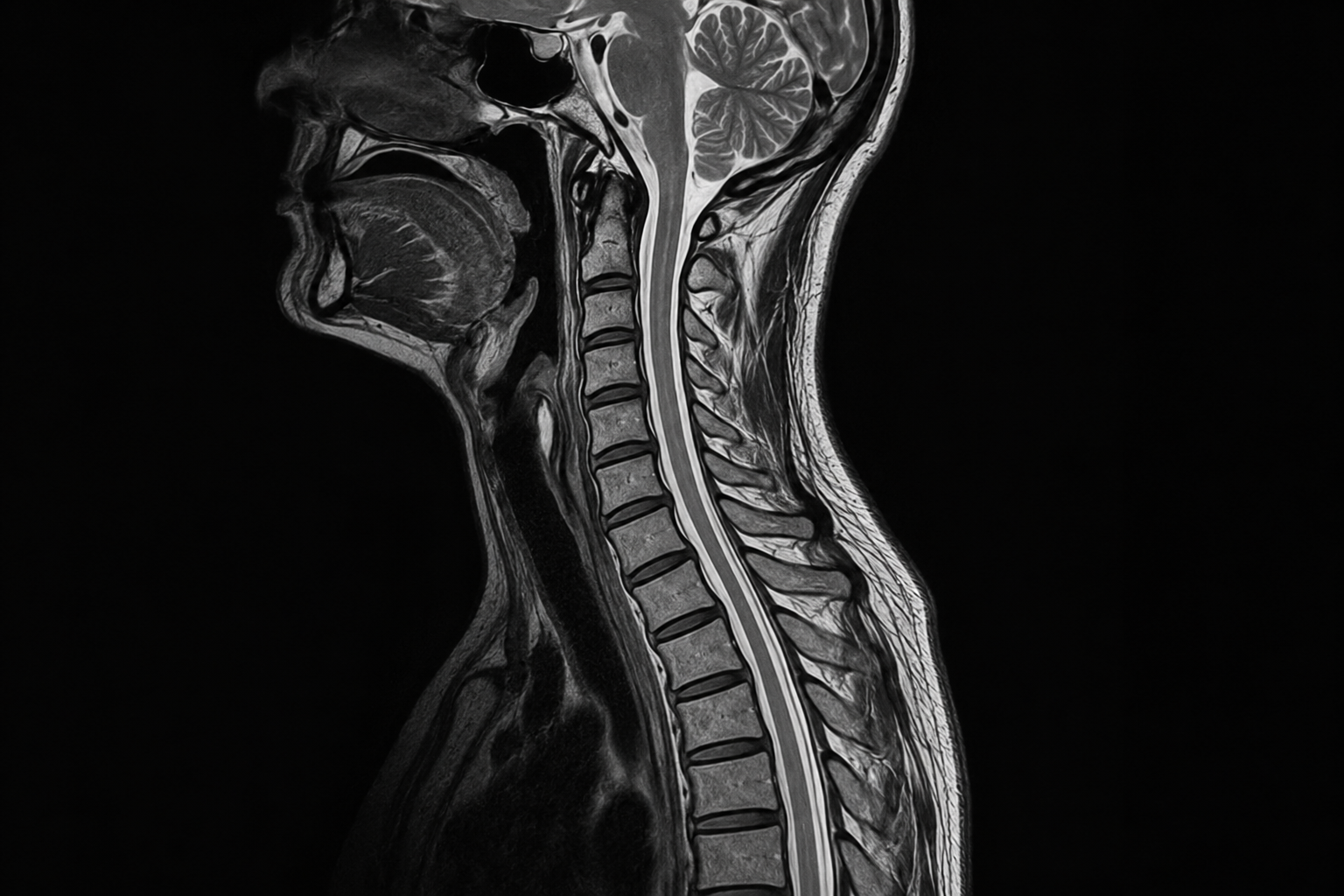
MRI is the key examination for discs, nerve roots, the spinal canal, spinal cord, bone marrow and surrounding soft tissues. It can show nerve compression, myelopathy, infection, inflammation and tumour. Contrast is added only when the clinical question requires it.
Routine imaging is not recommended for uncomplicated low back pain without red flags. Scans are most useful when the result is likely to change treatment.
05 · Treatment pathway
Start with the person, not the scan
For non-specific pain, care usually combines education, staying active within tolerance, exercise-based rehabilitation and appropriate pain management. Bed rest and fear of movement can delay recovery.
Surgical or specialist review is considered when there is significant neurological deficit, spinal-cord or cauda-equina compression, instability, infection, tumour, or disabling symptoms that persist despite suitable conservative care.
06 · Image-guided care
Targeted injections for selected patients
Periradicular therapy and transforaminal or epidural injections place medication close to an irritated nerve root or the epidural space under CT or fluoroscopic guidance. They may reduce inflammatory radicular pain and create a window for rehabilitation; they do not reverse every structural cause.
Selection depends on the clinical pattern, imaging, medicines and overall health. Benefits are usually temporary and variable. Bleeding, infection, dural puncture, allergic reaction, transient weakness and very rare neurological injury are discussed before treatment.
07 · Everyday spine health
Movement is part of prevention and recovery
Regular walking and strength, mobility and balance exercises support general and spinal health. Gradual progression, varied posture, sensible lifting and attention to sleep, smoking, body weight and bone health are more useful than searching for one perfect posture.
Persistent or recurrent symptoms deserve an individual plan. The goal is not simply a normal-looking scan, but safe function, confidence and participation in daily life.
08 · Evidence